
Article Figures & Data
Figures
- fig 1.
Inverse correlation between CNR of tumor-to-CSFventr on FLAIR images and ADC of epidermoid tumors in the corresponding cases (Pearson correlation coefficient, −.910; P = .0006)
- fig 2.
Right CPA epidermoid tumor in a 72-year-old man.
A-C, Contrast-enhanced T1-weighted (400/8/2 [TR/TE/excitations]) (A), fast T2-weighted (3000/98/2) (B) and proton density–weighted (3000/14/2) (C) images do not show the epidermoid tumor but only slight expansion of the arachnoid cistern in the right CPA (arrow).
D, Fast-FLAIR imaging (10002/148/1, inversion time of 2200) shows the tumor (arrows) as unevenly hyperintense relative to the CSF but hypointense relative to the brain.
E, Echo-planar DW imaging (1600/126 [TR/TE] with b value of 1000 s/mm2; field of view, 24 × 24 cm) reveals the tumor as a sharply hyperintense lesion (arrows) relative to the brain and CSF.
F, ADC map shows that the intensity of the tumor is similar to that of surrounding brain tissue but much different from that of CSF. Note the uneven diffusion in the lesion (arrows).
- fig 3.
Suprasellar region epidermoid tumor in a 47-year-old woman.
A, Fast-FLAIR imaging (10002/148/1, inversion time of 2200) shows that epidermoid tumor (black arrows) fills the suprasellar and right ambient cisterns; the hyperintensity in the left ambient and interpeduncular cisterns (white arrows) probably is caused by CSF flow artifacts.
B, Echo-planar DW imaging (1600/126 with b value of 1000 s/mm2; field of view, 36 × 24 cm) clearly shows that the tumor (black arrows) is in the suprasellar and right ambient cisterns; the signal in the left ambient and interpeduncular cisterns (white arrow) is greatly attenuated, indicating a fluid nature. Note the susceptibility artifacts (white arrowhead) at the anterior skull base.
C, ADC map shows that the ADC of the tumor (black arrows) is similar to that of surrounding brain tissue; the ADCs of left ambient and interpeduncular cisterns are similar to that of the eyes (white arrows).
- fig 4.
Right CPA epidermoid tumor in a 48-year-old man.
A, On fast-FLAIR imaging (10002/148/1, inversion time of 2200), epidermoid tumor appears to extend over the midline in the prepontine cistern (arrow).
B, Echo-planar DW imaging (1600/126 with b value of 1000 s/mm2; field of view, 36 × 24 cm) reveals that the lesion (black arrow) is limited in the right CPA. Note the susceptibility artifacts near the temporal bone (white arrows).
C, ADC map shows that the tumor (arrow) has an ADC similar to that of surrounding brain tissue.


Tables
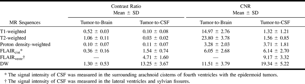
 TABLE 1:
Contrast ratios, CNRs of tumor-to-brain and tumor-to-CSF in epidermoid tumors
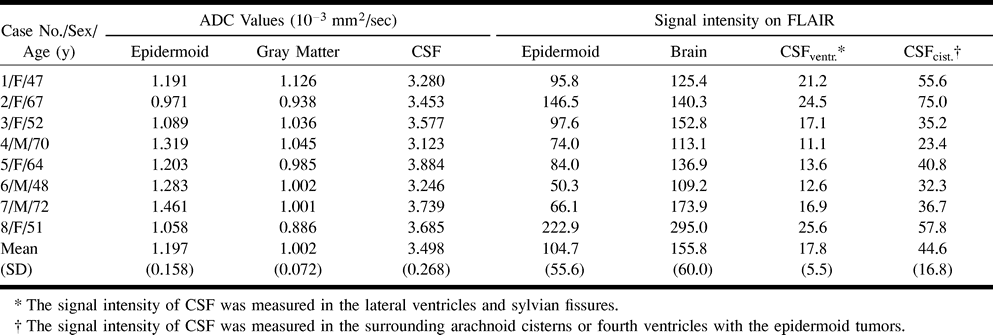
- TABLE 2:
Mean ADC Values and signal intensity on FLAIR of epidermoid tumor, brain tissue, and CSF
In this issue


Jump to section
Related Articles
Cited By...
- Diffusion Analysis of Intracranial Epidermoid, Head and Neck Epidermal Inclusion Cyst, and Temporal Bone Cholesteatoma
- Atypical presentation of large intracranial epidermoid tumour in a child
- Apparent Diffusion Coefficient Values of Middle Ear Cholesteatoma Differ from Abscess and Cholesteatoma Admixed Infection
- Imaging Lesions of the Cavernous Sinus