
Article Figures & Data
Figures
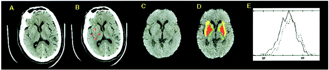
- fig 1.
Large left MCA stroke (patient case 1).
A, Raw CT scan shows left MCA infarct predominantly involving the insula, with marginal involvement of the lentiform nucleus (right of image is left of patient).
B, CT scan after automated scalp stripping and normalization to Talairach atlas (right of image is right of patient).
C, Automated segmentation color coded onto normalized CT scan shows excellent registration of lentiform nucleus (red) and insular cortex (yellow). Internal capsule is displayed in green.
D, Histogram plot for lentiform nucleus shows mild leftward shift for the left lentiform nucleus (dotted line, P < .0196 using a Wilcoxon statistic corrected for spatial autocorrelation).
E, Histogram plot for insula shows a large leftward shift (P < 1 × 10⁻⁹).
- fig 2.
Subtle right MCA stroke (patient case 5).
A, Raw CT scan shows subtle right MCA territory infarct (right of image is left of patient) involving the insula and posterior putamen.
B, Posterior putamen and insula are highlighted.
C, CT scan after automated scalp stripping and normalization to Talairach atlas (right of image is right of patient).
D, Automated segmentation color coded onto normalized CT scan (as in fig 1) shows excellent registration of basal ganglia structures and insular cortex.
E, Histogram plot for insula shows leftward shift for the right insula (solid line, P < 1 × 10⁻⁹).


