
Article Figures & Data
Figures
- fig 1.
A 55-year-old man (patient 1) with a visual field defect.
A, Visual field examination shows right homonymous inferior quadrantanopsia.
B, T2-weighted image [4500/120 (TR/TE)] shows infarction in the left occipital lobe (arrow). The left primary visual cortex itself and the visual pathway are spared.
C, Digital subtraction angiogram shows focal stenosis at the left posterior cerebral artery (arrows).
D, fMR [90/56/40° (TR/TE/flip angle)] shows decreased activity in the left visual cortex.
- fig 2.
A 52-year-old woman (patient 2) with several previous episodes of visual disturbance.
A and B, Visual field examination (A) and T2-weighted image [4500/120 (TR/TE)] (B) are normal.
C, MR angiogram [30/6.4/25° (TR/TE/flip angle)] shows poor blood flow (arrowheads) in the right posterior cerebral artery.
D, fMR [90/56/40° (TR/TE/flip angle)] shows decreased activity in the right visual cortex.
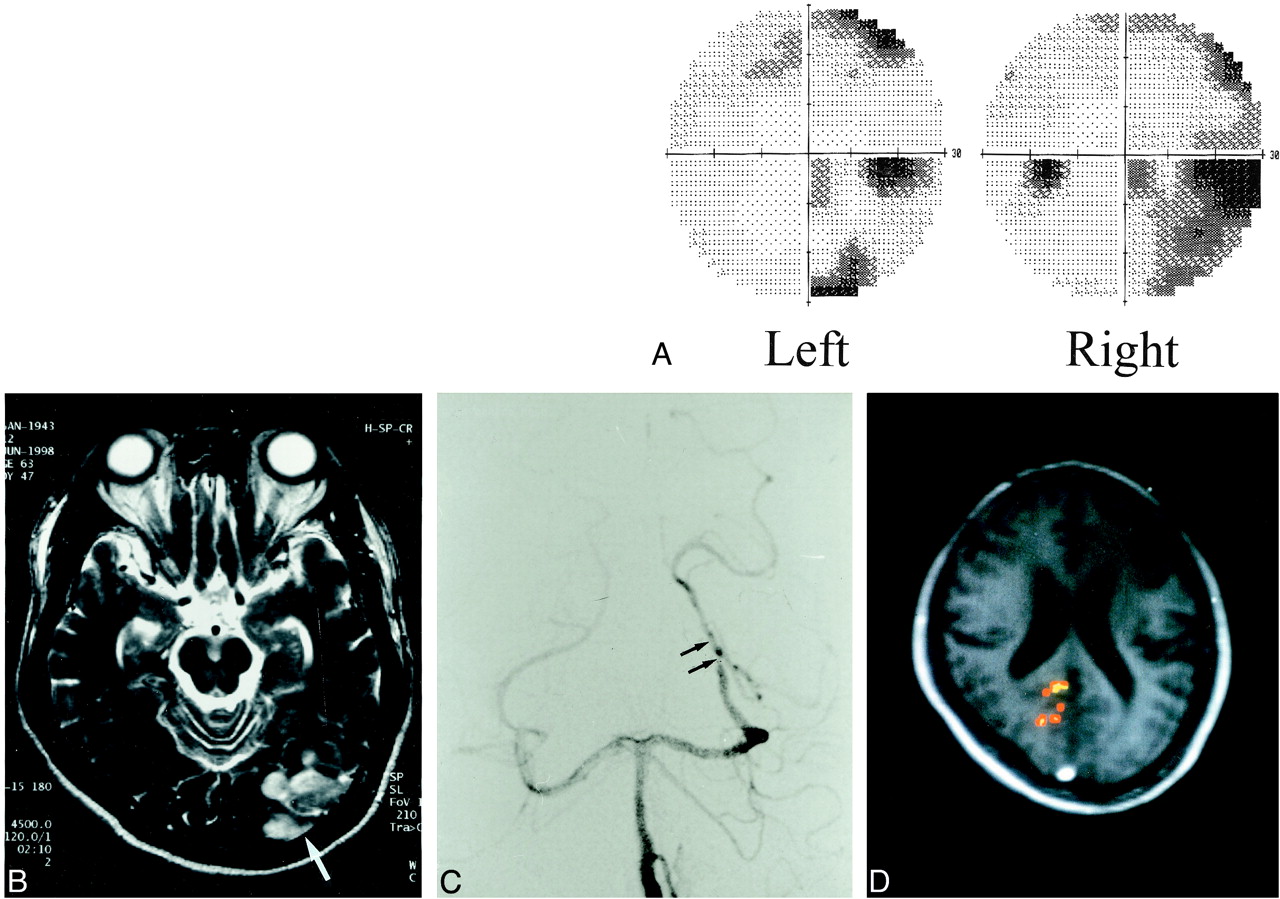
- fig 3.
A 29-year-old woman (patient 3) with intermittent left homonymous hemianopsia.
A, T2-weighted image [4500/120 (TR/TE)] is normal.
B and C, Digital subtraction angiogram (B) shows discontinuity of the right vertebral artery and collateral vessels (arrow). The clinical and angiographic diagnosis was dissection of the vertebrobasilar artery. The right posterior communicating artery was hypoplastic (C), whereas the left posterior communicating artery (arrow) showed good patency. The clinical symptom of visual problem in this patient was believed to appear when blood flow from the basilar artery decreased.
D, fMR [90/56/40° (TR/TE/flip angle)] shows decreased activity in the right visual cortex.


Tables
 TABLE 1:
Summary of the clinical findings and results of fMRI, visual field examination, conventional MR imaging, and vascular imaging
- TABLE 2:
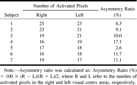
Number of activated pixels and asymmetry ratios in the normal control group
- TABLE 3:
Number of activated pixels and asymmetry ratios in the patient group


