Advertisement

Main menu

  • Home
  • Content
    • Current Issue
    • Accepted Manuscripts
    • Article Preview
    • Past Issue Archive
    • Video Articles
    • AJNR Case Collection
    • Case of the Week Archive
    • Case of the Month Archive
    • Classic Case Archive
  • Special Collections
    • AJNR Awards
    • Low-Field MRI
    • Alzheimer Disease
    • ASNR Foundation Special Collection
    • Photon-Counting CT
    • View All
  • Multimedia
    • AJNR Podcasts
    • AJNR SCANtastic
    • Trainee Corner
    • MRI Safety Corner
    • Imaging Protocols
  • For Authors
    • Submit a Manuscript
    • Submit a Video Article
    • Submit an eLetter to the Editor/Response
    • Manuscript Submission Guidelines
    • Statistical Tips
    • Fast Publishing of Accepted Manuscripts
    • Graphical Abstract Preparation
    • Imaging Protocol Submission
    • Author Policies
  • About Us
    • About AJNR
    • Editorial Board
    • Editorial Board Alumni
  • More
    • Become a Reviewer/Academy of Reviewers
    • Subscribers
    • Permissions
    • Alerts
    • Feedback
    • Advertisers
    • ASNR Home

User menu

  • Alerts
  • Log in

Search

  • Advanced search
American Journal of Neuroradiology

American Journal of Neuroradiology

ASHNR American Society of Functional Neuroradiology ASHNR American Society of Pediatric Neuroradiology ASSR
  • Alerts
  • Log in

Advanced Search

  • Home
  • Content
    • Current Issue
    • Accepted Manuscripts
    • Article Preview
    • Past Issue Archive
    • Video Articles
    • AJNR Case Collection
    • Case of the Week Archive
    • Case of the Month Archive
    • Classic Case Archive
  • Special Collections
    • AJNR Awards
    • Low-Field MRI
    • Alzheimer Disease
    • ASNR Foundation Special Collection
    • Photon-Counting CT
    • View All
  • Multimedia
    • AJNR Podcasts
    • AJNR SCANtastic
    • Trainee Corner
    • MRI Safety Corner
    • Imaging Protocols
  • For Authors
    • Submit a Manuscript
    • Submit a Video Article
    • Submit an eLetter to the Editor/Response
    • Manuscript Submission Guidelines
    • Statistical Tips
    • Fast Publishing of Accepted Manuscripts
    • Graphical Abstract Preparation
    • Imaging Protocol Submission
    • Author Policies
  • About Us
    • About AJNR
    • Editorial Board
    • Editorial Board Alumni
  • More
    • Become a Reviewer/Academy of Reviewers
    • Subscribers
    • Permissions
    • Alerts
    • Feedback
    • Advertisers
    • ASNR Home
  • Follow AJNR on Twitter
  • Visit AJNR on Facebook
  • Follow AJNR on Instagram
  • Join AJNR on LinkedIn
  • RSS Feeds

AJNR Awards, New Junior Editors, and more. Read the latest AJNR updates

Research ArticleBRAIN

Initial and Follow-up MR Imaging Findings in AIDS-Related Progressive Multifocal Leukoencephalopathy Treated with Highly Active Antiretroviral Therapy

Majda M. Thurnher, M. Judith Donovan Post, Armin Rieger, Christina Kleibl-Popov, Christian Loewe and Erwin Schindler
American Journal of Neuroradiology May 2001, 22 (5) 977-984;
Majda M. Thurnher
  • Find this author on Google Scholar
  • Find this author on PubMed
  • Search for this author on this site
M. Judith Donovan Post
  • Find this author on Google Scholar
  • Find this author on PubMed
  • Search for this author on this site
Armin Rieger
  • Find this author on Google Scholar
  • Find this author on PubMed
  • Search for this author on this site
Christina Kleibl-Popov
  • Find this author on Google Scholar
  • Find this author on PubMed
  • Search for this author on this site
Christian Loewe
  • Find this author on Google Scholar
  • Find this author on PubMed
  • Search for this author on this site
Erwin Schindler
  • Find this author on Google Scholar
  • Find this author on PubMed
  • Search for this author on this site
  • Article
  • Figures & Data
  • Info & Metrics
  • Responses
  • References
  • PDF
Loading

Article Figures & Data

Figures

  • Tables
  • fig 1.
    • Download figure
    • Open in new tab
    • Download powerpoint
    fig 1.

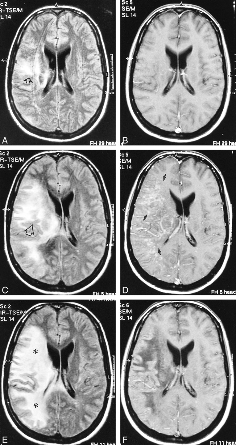
    Case 2: 32-year-old HIV-positive woman with PML and survival time exceeding 22 months.

    A and B, Initial MR study, December 1998. Axial FLAIR-FSE (TR/TE = 10000/150, TI = 2600) image (A) shows high-signal-intensity lesion in the white matter of the right centrum semiovale (arrow). There is no mass effect. No enhancement was present on contrast-enhanced T1-weighted (550/20) image (B).

    C and D, Follow-up MR study, January 1999 (1 month after initiation of HAART). FLAIR-FSE (10000/150, TI = 2600) image (C) shows progression of white matter abnormalities (arrow) with mass effect and compression of right ventricle. Increased hypoattenuation of the lesion was evident on T1-weighted (550/20) images (not shown). On contrast-enhanced T1-weighted (550/20) image (D), diffuse enhancement of white matter abnormalities is present (arrows).

    E and F, Subsequent MR study, February 1999 (2 months after initiation of HAART). FLAIR-FSE (10000/150, TI = 2600) image (E) shows further progression of the high-signal-intensity lesions of white matter disease (asterisks). Slight enhancement is still present on contrast-enhanced T1-weighted (550/20) image (F). Note increased hypoattenuation of the lesions and regression of the mass effect. Continued on page 982
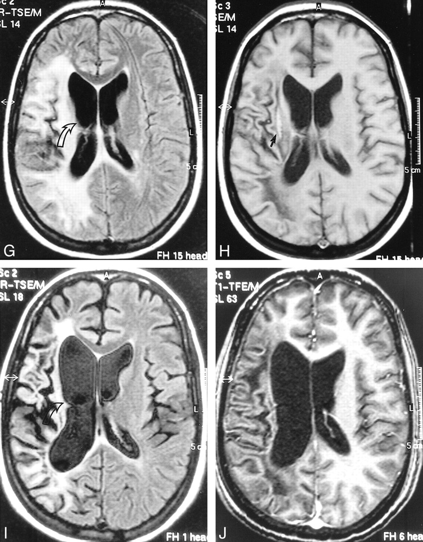
  • fig 1.
    • Download figure
    • Open in new tab
    • Download powerpoint
    fig 1.

    Continued.

    G and H, Further follow-up MR study, April 1999 (4 months after initiation of HAART). FLAIR-FSE (10000/150, TI = 2600) image (G) reveals regression of the white matter changes with partially present low signal (corresponding to areas of increased hypoattenuation on T1-weighted images). Note atrophic changes of the right hemisphere with widening of the ventricle (arrow). Further increase in hypointensity is evident on T1-weighted (550/20) image (H). Elongated hyperintense area is seen medial to the sylvian fissure, representing subacute hemorrhage (arrow). Enhancement was not present on contrast-enhanced T1-weighted image (not shown).

    I and J, Follow-up MR examination, September 2000 (21 months after initiation of HAART). Axial FLAIR-FSE (7384/130, TI = 2100) image (I) and contrast-enhanced T1-weighted (8/2.7, flip angle = 10°) image (J) show atrophic changes in the right hemisphere with widening of the sulci and further ex-vacuo widening of the right ventricle (arrow). Note low signal on the FLAIR-FSE image, representing leukomalacia

  • fig 2.
    • Download figure
    • Open in new tab
    • Download powerpoint
    fig 2.

    Case 4: 28-year-old man with AIDS who presented with psychomotor slowing and hemianopsia. Stereotactic biopsy revealed PML.

    A–C, Initial MR study, March 1998. Axial FLAIR-FSE (10000/150, TI = 2600) image (A) shows scalloped, high-signal-intensity lesion in the parietooccipital white matter with extension to dorsal part of the corpus callosum on both sides (arrow). There is no mass effect. The lesion is hypointense on noncontrast T1-weighted SE (550/20) image (B). No enhancement is observed on contrast-enhanced T1-weighted SE (550/20) image (C).

    D–F, Follow-up MR study, May 1998 (4 weeks after the start of HAART). FLAIR-FSE (10000/150, TI = 2600) image (D) shows progression of the white matter lesion (arrow) with involvement of the right parietal white matter and contralateral occipital white matter. Increased hypointensity is evident on noncontrast T1-weighted (550/20) image (E). Contrast-enhanced T1-weighted (550/20) image shows no enhancement (F)

Tables

  • Figures
  • TABLE 1:
    • View popup
    • Download powerpoint
    TABLE 1:

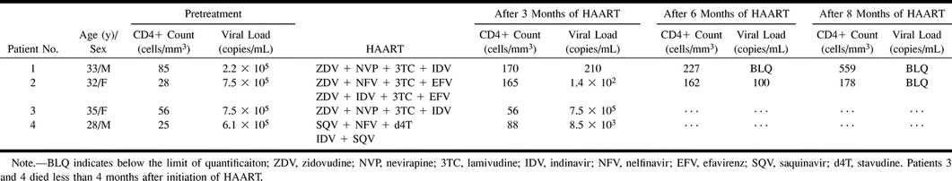
    Immunologic and virologic values in four patients with AIDS-associated PML receiving HAART

  • TABLE 2:
    • View popup
    • Download powerpoint
    TABLE 2:

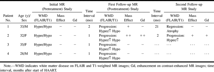
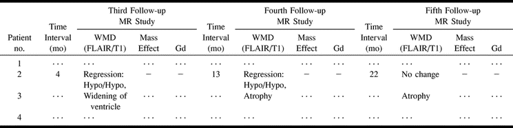
    Initial and follow-up MR findings in four patients with AIDS-associated PML receiving HAART

  • TABLE 2:
    • View popup
    • Download powerpoint
    TABLE 2:

    Continued

PreviousNext
Back to top

In this issue

American Journal of Neuroradiology
Vol. 22, Issue 5
1 May 2001
  • Table of Contents
  • Index by author
Advertisement
Print
Download PDF
Email Article

Thank you for your interest in spreading the word on American Journal of Neuroradiology.

NOTE: We only request your email address so that the person you are recommending the page to knows that you wanted them to see it, and that it is not junk mail. We do not capture any email address.

Enter multiple addresses on separate lines or separate them with commas.
Initial and Follow-up MR Imaging Findings in AIDS-Related Progressive Multifocal Leukoencephalopathy Treated with Highly Active Antiretroviral Therapy
(Your Name) has sent you a message from American Journal of Neuroradiology
(Your Name) thought you would like to see the American Journal of Neuroradiology web site.
CAPTCHA
This question is for testing whether or not you are a human visitor and to prevent automated spam submissions.
Cite this article
Majda M. Thurnher, M. Judith Donovan Post, Armin Rieger, Christina Kleibl-Popov, Christian Loewe, Erwin Schindler
Initial and Follow-up MR Imaging Findings in AIDS-Related Progressive Multifocal Leukoencephalopathy Treated with Highly Active Antiretroviral Therapy
American Journal of Neuroradiology May 2001, 22 (5) 977-984;

Citation Manager Formats

  • BibTeX
  • Bookends
  • EasyBib
  • EndNote (tagged)
  • EndNote 8 (xml)
  • Medlars
  • Mendeley
  • Papers
  • RefWorks Tagged
  • Ref Manager
  • RIS
  • Zotero
0 Responses
Respond to this article
Share
Bookmark this article
Initial and Follow-up MR Imaging Findings in AIDS-Related Progressive Multifocal Leukoencephalopathy Treated with Highly Active Antiretroviral Therapy
Majda M. Thurnher, M. Judith Donovan Post, Armin Rieger, Christina Kleibl-Popov, Christian Loewe, Erwin Schindler
American Journal of Neuroradiology May 2001, 22 (5) 977-984;
del.icio.us logo Twitter logo Facebook logo Mendeley logo
  • Tweet Widget
  • Facebook Like
  • Google Plus One

Jump to section

  • Article
    • Abstract
    • Methods
    • Results
    • Discussion
    • Conclusion
    • Footnotes
    • References
  • Figures & Data
  • Info & Metrics
  • Responses
  • References
  • PDF

Related Articles

  • No related articles found.
  • PubMed
  • Google Scholar

Cited By...

  • Susceptibility-Weighted MR Imaging Hypointense Rim in Progressive Multifocal Leukoencephalopathy: The End Point of Neuroinflammation and a Potential Outcome Predictor
  • CNS-Immune Reconstitution Inflammatory Syndrome in the Setting of HIV Infection, Part 1: Overview and Discussion of Progressive Multifocal Leukoencephalopathy-Immune Reconstitution Inflammatory Syndrome and Cryptococcal-Immune Reconstitution Inflammatory Syndrome
  • JC Virus Infection of the Brain
  • Immune Reconstitution Inflammatory Syndrome and Cerebral Toxoplasmosis
  • Use of Diffusion-Weighted Imaging to Evaluate the Initial Response of Progressive Multifocal Leukoencephalopathy to Highly Active Antiretroviral Therapy: Early Experience
  • Natalizumab and progressive multifocal leucoencephalopathy
  • Crossref
  • Google Scholar

This article has not yet been cited by articles in journals that are participating in Crossref Cited-by Linking.

More in this TOC Section

  • Multimodal CT Provides Improved Performance for Lacunar Infarct Detection
  • Optimal MRI Sequence for Identifying Occlusion Location in Acute Stroke: Which Value of Time-Resolved Contrast-Enhanced MRA?
  • Evaluating the Effects of White Matter Multiple Sclerosis Lesions on the Volume Estimation of 6 Brain Tissue Segmentation Methods
Show more BRAIN

Similar Articles

Advertisement

Indexed Content

  • Current Issue
  • Accepted Manuscripts
  • Article Preview
  • Past Issues
  • Editorials
  • Editor's Choice
  • Fellows' Journal Club
  • Letters to the Editor
  • Video Articles

Cases

  • Case Collection
  • Archive - Case of the Week
  • Archive - Case of the Month
  • Archive - Classic Case

More from AJNR

  • Trainee Corner
  • Imaging Protocols
  • MRI Safety Corner

Multimedia

  • AJNR Podcasts
  • AJNR Scantastics

Resources

  • Turnaround Time
  • Submit a Manuscript
  • Submit a Video Article
  • Submit an eLetter to the Editor/Response
  • Manuscript Submission Guidelines
  • Statistical Tips
  • Fast Publishing of Accepted Manuscripts
  • Graphical Abstract Preparation
  • Imaging Protocol Submission
  • Evidence-Based Medicine Level Guide
  • Publishing Checklists
  • Author Policies
  • Become a Reviewer/Academy of Reviewers
  • News and Updates

About Us

  • About AJNR
  • Editorial Board
  • Editorial Board Alumni
  • Alerts
  • Permissions
  • Not an AJNR Subscriber? Join Now
  • Advertise with Us
  • Librarian Resources
  • Feedback
  • Terms and Conditions
  • AJNR Editorial Board Alumni

American Society of Neuroradiology

  • Not an ASNR Member? Join Now

© 2025 by the American Society of Neuroradiology All rights, including for text and data mining, AI training, and similar technologies, are reserved.
Print ISSN: 0195-6108 Online ISSN: 1936-959X

Powered by HighWire