Advertisement

Main menu

  • Home
  • Content
    • Current Issue
    • Accepted Manuscripts
    • Article Preview
    • Past Issue Archive
    • Video Articles
    • AJNR Case Collection
    • Case of the Week Archive
    • Case of the Month Archive
    • Classic Case Archive
  • Special Collections
    • AJNR Awards
    • Low-Field MRI
    • Alzheimer Disease
    • ASNR Foundation Special Collection
    • Photon-Counting CT
    • View All
  • Multimedia
    • AJNR Podcasts
    • AJNR SCANtastic
    • Trainee Corner
    • MRI Safety Corner
    • Imaging Protocols
  • For Authors
    • Submit a Manuscript
    • Submit a Video Article
    • Submit an eLetter to the Editor/Response
    • Manuscript Submission Guidelines
    • Statistical Tips
    • Fast Publishing of Accepted Manuscripts
    • Graphical Abstract Preparation
    • Imaging Protocol Submission
    • Author Policies
  • About Us
    • About AJNR
    • Editorial Board
    • Editorial Board Alumni
  • More
    • Become a Reviewer/Academy of Reviewers
    • Subscribers
    • Permissions
    • Alerts
    • Feedback
    • Advertisers
    • ASNR Home

User menu

  • Alerts
  • Log in

Search

  • Advanced search
American Journal of Neuroradiology

American Journal of Neuroradiology

ASHNR American Society of Functional Neuroradiology ASHNR American Society of Pediatric Neuroradiology ASSR
  • Alerts
  • Log in

Advanced Search

  • Home
  • Content
    • Current Issue
    • Accepted Manuscripts
    • Article Preview
    • Past Issue Archive
    • Video Articles
    • AJNR Case Collection
    • Case of the Week Archive
    • Case of the Month Archive
    • Classic Case Archive
  • Special Collections
    • AJNR Awards
    • Low-Field MRI
    • Alzheimer Disease
    • ASNR Foundation Special Collection
    • Photon-Counting CT
    • View All
  • Multimedia
    • AJNR Podcasts
    • AJNR SCANtastic
    • Trainee Corner
    • MRI Safety Corner
    • Imaging Protocols
  • For Authors
    • Submit a Manuscript
    • Submit a Video Article
    • Submit an eLetter to the Editor/Response
    • Manuscript Submission Guidelines
    • Statistical Tips
    • Fast Publishing of Accepted Manuscripts
    • Graphical Abstract Preparation
    • Imaging Protocol Submission
    • Author Policies
  • About Us
    • About AJNR
    • Editorial Board
    • Editorial Board Alumni
  • More
    • Become a Reviewer/Academy of Reviewers
    • Subscribers
    • Permissions
    • Alerts
    • Feedback
    • Advertisers
    • ASNR Home
  • Follow AJNR on Twitter
  • Visit AJNR on Facebook
  • Follow AJNR on Instagram
  • Join AJNR on LinkedIn
  • RSS Feeds

AJNR Awards, New Junior Editors, and more. Read the latest AJNR updates

Research ArticleHead and Neck Imaging

Mycotic Aneurysm and Cerebral Infarction Resulting from Fungal Sinusitis: Imaging and Pathologic Correlation

Robert W. Hurst, Alex Judkins, William Bolger, Albert Chu and Laurie A. Loevner
American Journal of Neuroradiology May 2001, 22 (5) 858-863;
Robert W. Hurst
  • Find this author on Google Scholar
  • Find this author on PubMed
  • Search for this author on this site
Alex Judkins
  • Find this author on Google Scholar
  • Find this author on PubMed
  • Search for this author on this site
William Bolger
  • Find this author on Google Scholar
  • Find this author on PubMed
  • Search for this author on this site
Albert Chu
  • Find this author on Google Scholar
  • Find this author on PubMed
  • Search for this author on this site
Laurie A. Loevner
  • Find this author on Google Scholar
  • Find this author on PubMed
  • Search for this author on this site
  • Article
  • Figures & Data
  • Info & Metrics
  • Responses
  • References
  • PDF
Loading

Article Figures & Data

Figures

  • fig 1.
    • Download figure
    • Open in new tab
    • Download powerpoint
    fig 1.

    A and B, Coronal CT scan shows soft tissue density within the sphenoid sinus, with bony destruction in the region of the cavernous portion of the right internal carotid artery

  • fig 2.
    • Download figure
    • Open in new tab
    • Download powerpoint
    fig 2.

    A, Endoscopic view of the sphenoid sinus following removal of the inspissated mucus and fungal debris. The most lateral and inferior portions of the sinus are evident (arrowhead). The aneurysm (a) projects from the superior aspect of the sphenoid, to nearly fill the sinus (s = midline nasal septum).

    B, Endoscopic view of the sphenoid sinus approximately 5 days after embolization. There is no evidence of recurrent fungal debris and the embolized aneurysm is much smaller
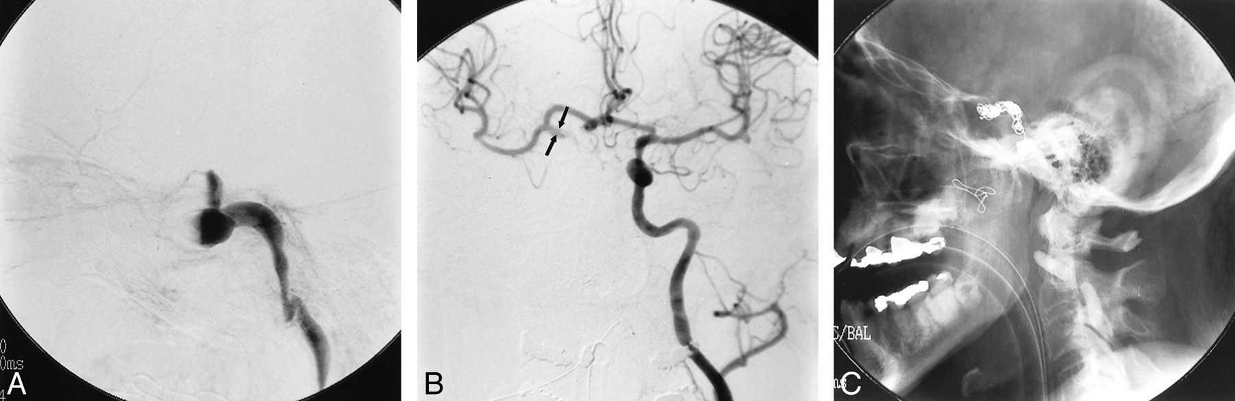
  • fig 3.
    • Download figure
    • Open in new tab
    • Download powerpoint
    fig 3.

    A, Lateral view, angiogram of the right internal carotid artery following initial debridement and packing of the sphenoid sinus shows fusiform dilation of the cavernous portion of the vessel. Occlusion of the carotid artery resulted from packing of the sphenoid sinus at the time of surgery.

    B, Injection of the left internal carotid artery shows good filling across the anterior communicating artery. The visualized portions of the supraclinoid right internal carotid artery (arrows) and the right middle cerebral artery are of normal caliber.

    C, Postembolization plain film shows microcoils within the lumen of the aneurysm and proximal detachable balloons occluding the right internal carotid artery
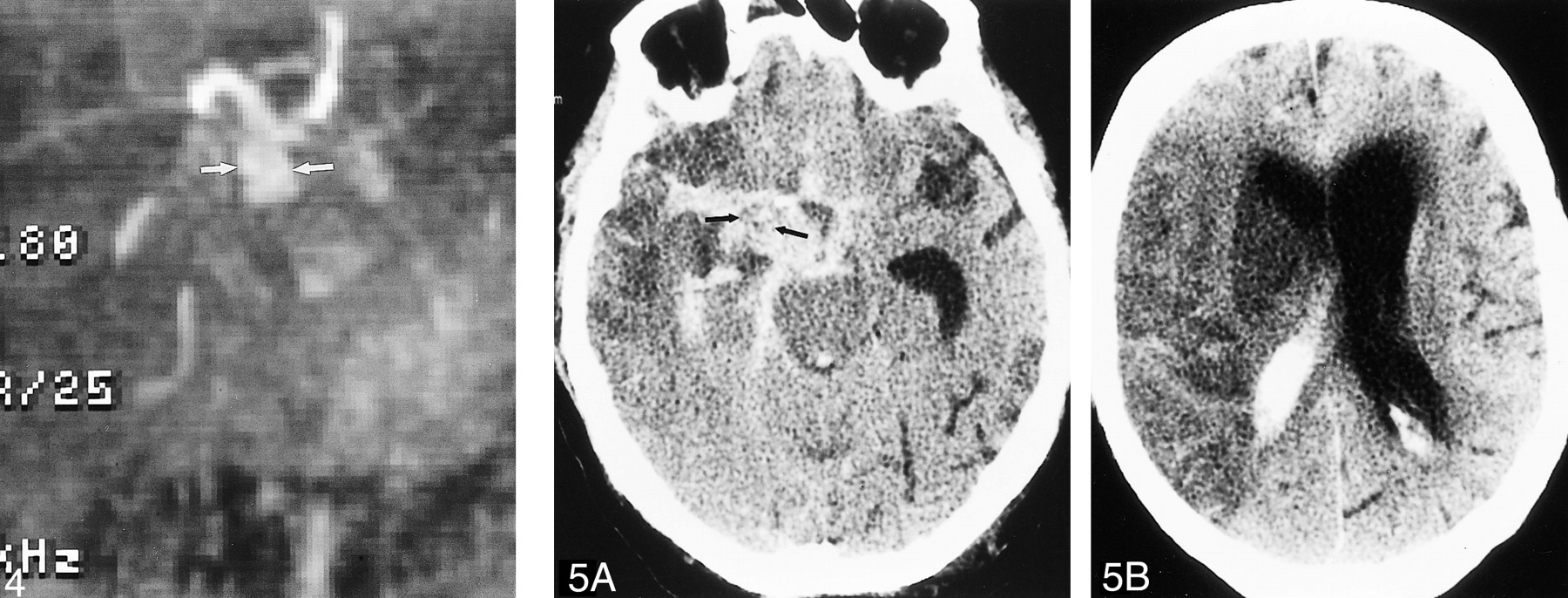
  • fig 4.
    • Download figure
    • Open in new tab
    • Download powerpoint
    fig 4.

    MR angiogram (time of flight, 48/5.9 [TR/TE]) shows dilated supraclinoid right internal carotid artery (arrows) representing aneurysmal progression from angiogram performed at the time of embolization.

    fig 5. A, Unenhanced CT scan shows extensive subarachnoid hemorrhage outlining soft tissue mass (arrows) in the right basilar cistern, which represents the supraclinoid portion of the aneurysm.

    B, Hypodense cerebral infarct with mass effect is present throughout the right middle cerebral artery distribution

  • fig 6.
    • Download figure
    • Open in new tab
    • Download powerpoint
    fig 6.

    A, Aneurysmal dilation of the supraclinoid segment of the right internal carotid artery. Rupture sites (arrows) are present between the posterior communicating and anterior choroidal arteries, as well as along the M1 segment of the right middle cerebral artery.

    B, Histopathologic examination shows severe inflammatory change, necrosis of the arterial wall and elastica, intralumenal thrombosis, and focal rupture site (asterisk). Inset: (Grocott stain) Septated hyphae branching at acute angles, morphologically consistent with aspergillus species, located predominantely in the vessel wall and adventitia are seen

PreviousNext
Back to top

In this issue

American Journal of Neuroradiology
Vol. 22, Issue 5
1 May 2001
  • Table of Contents
  • Index by author
Advertisement
Print
Download PDF
Email Article

Thank you for your interest in spreading the word on American Journal of Neuroradiology.

NOTE: We only request your email address so that the person you are recommending the page to knows that you wanted them to see it, and that it is not junk mail. We do not capture any email address.

Enter multiple addresses on separate lines or separate them with commas.
Mycotic Aneurysm and Cerebral Infarction Resulting from Fungal Sinusitis: Imaging and Pathologic Correlation
(Your Name) has sent you a message from American Journal of Neuroradiology
(Your Name) thought you would like to see the American Journal of Neuroradiology web site.
CAPTCHA
This question is for testing whether or not you are a human visitor and to prevent automated spam submissions.
Cite this article
Robert W. Hurst, Alex Judkins, William Bolger, Albert Chu, Laurie A. Loevner
Mycotic Aneurysm and Cerebral Infarction Resulting from Fungal Sinusitis: Imaging and Pathologic Correlation
American Journal of Neuroradiology May 2001, 22 (5) 858-863;

Citation Manager Formats

  • BibTeX
  • Bookends
  • EasyBib
  • EndNote (tagged)
  • EndNote 8 (xml)
  • Medlars
  • Mendeley
  • Papers
  • RefWorks Tagged
  • Ref Manager
  • RIS
  • Zotero
0 Responses
Respond to this article
Share
Bookmark this article
Mycotic Aneurysm and Cerebral Infarction Resulting from Fungal Sinusitis: Imaging and Pathologic Correlation
Robert W. Hurst, Alex Judkins, William Bolger, Albert Chu, Laurie A. Loevner
American Journal of Neuroradiology May 2001, 22 (5) 858-863;
del.icio.us logo Twitter logo Facebook logo Mendeley logo
  • Tweet Widget
  • Facebook Like
  • Google Plus One

Jump to section

  • Article
    • Abstract
    • Case Report
    • Discussion
    • Conclusion
    • Footnotes
    • References
  • Figures & Data
  • Info & Metrics
  • Responses
  • References
  • PDF

Related Articles

  • No related articles found.
  • PubMed
  • Google Scholar

Cited By...

  • Disseminated cryptococcosis in an apparently immunocompetent patient presenting with primary intraventricular haemorrhage
  • Retrospective Review of Cerebral Mycotic Aneurysms in 26 Patients: Focus on Treatment in Strongly Immunocompromised Patients with a Brief Literature Review
  • Endovascular Treatment of Epistaxis
  • Crossref
  • Google Scholar

This article has not yet been cited by articles in journals that are participating in Crossref Cited-by Linking.

More in this TOC Section

  • FDG Uptake and Thyroid Cancer Post-BRAF Therapy
  • Normal Facial Nerve Enhancement on VIBE Sequence
  • Lipochoristomas and inner ear signal
Show more Head and Neck Imaging

Similar Articles

Advertisement

Indexed Content

  • Current Issue
  • Accepted Manuscripts
  • Article Preview
  • Past Issues
  • Editorials
  • Editor's Choice
  • Fellows' Journal Club
  • Letters to the Editor
  • Video Articles

Cases

  • Case Collection
  • Archive - Case of the Week
  • Archive - Case of the Month
  • Archive - Classic Case

More from AJNR

  • Trainee Corner
  • Imaging Protocols
  • MRI Safety Corner

Multimedia

  • AJNR Podcasts
  • AJNR Scantastics

Resources

  • Turnaround Time
  • Submit a Manuscript
  • Submit a Video Article
  • Submit an eLetter to the Editor/Response
  • Manuscript Submission Guidelines
  • Statistical Tips
  • Fast Publishing of Accepted Manuscripts
  • Graphical Abstract Preparation
  • Imaging Protocol Submission
  • Evidence-Based Medicine Level Guide
  • Publishing Checklists
  • Author Policies
  • Become a Reviewer/Academy of Reviewers
  • News and Updates

About Us

  • About AJNR
  • Editorial Board
  • Editorial Board Alumni
  • Alerts
  • Permissions
  • Not an AJNR Subscriber? Join Now
  • Advertise with Us
  • Librarian Resources
  • Feedback
  • Terms and Conditions
  • AJNR Editorial Board Alumni

American Society of Neuroradiology

  • Not an ASNR Member? Join Now

© 2025 by the American Society of Neuroradiology All rights, including for text and data mining, AI training, and similar technologies, are reserved.
Print ISSN: 0195-6108 Online ISSN: 1936-959X

Powered by HighWire