
Article Figures & Data
Figures
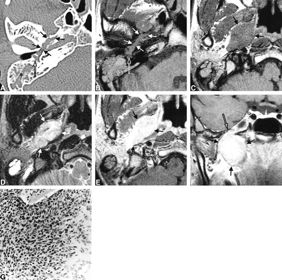
- fig 1.
35-year-old man with a 7-month history of right tinnitus and hearing disturbance.
A, Axial high-resolution CT scan shows tumor extension into the middle ear (open arrow) through the eustachian tube and widening of the bony eustachian tube (closed arrows). Mastoid opacification (asterisk) is secondary to eustachian tube obstruction.
B, Axial T1-weighted MR image (550/20/2 [TR/TE/excitations]) at the same level as A shows tumor filling the eustachian tube (closed arrows) and middle ear cavity (open arrow), extending from the parapharyngeal tumor (asterisk).
C, Axial T1-weighted MR image (550/20/2) shows an ovoid homogeneous mass (arrows) in the right parapharyngeal space isointense with surrounding muscle. The mass has a well-demarcated margin and displaces parapharyngeal fat laterally (asterisk).
D, Axial T2-weighted MR image (4858/112/4) at the same level as B shows slightly hyperintense mass (arrows) in the parapharyngeal space.
E, Contrast-enhanced axial T1-weighted MR image (550/20/2) shows homogeneously intense enhancement of the parapharyngeal mass (arrows).
F, Contrast-enhanced coronal T1-weighted MR image (722/20/2) shows the enhancing mass in the parapharyngeal space. Note close relationship between the mass (closed arrows) and the foramen ovale (open arrow), which is widened.
G, Photomicrograph of the excised mass. The mass is made up of relatively hypo- and hypercellular areas. The cells are spindle- or polyhedral-shaped and show moderate degrees of cellular pleomorphism and frequent mitoses (hematoxylin-eosin, original magnification ×100).


