
Article Figures & Data
Figures
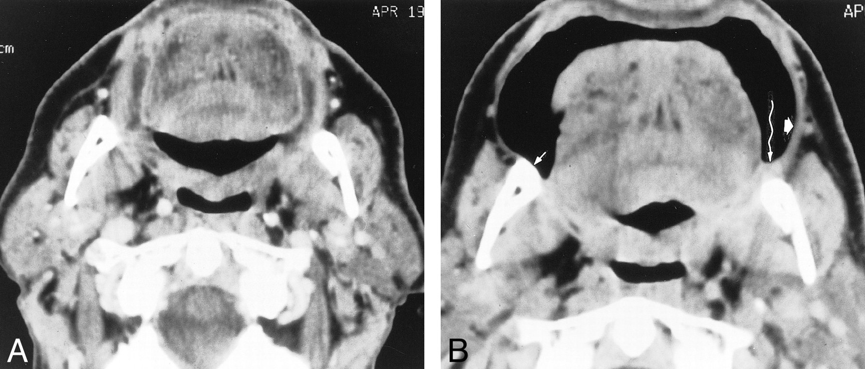
- fig 1.
Normal anatomy on puffed-cheek CT scans in two patients. A, 42-year-old woman with cheek swelling. Puffed-cheek axial CT scan at the level of the maxilla is normal. The patient's physical examination and subsequent follow-up were also normal. The muscles of facial expression, seen especially well on the puffed-cheek CT scan, include the orbicularis oris (long arrows), levator anguli oris (open white arrows), risorius, and buccinator. Note streak artifacts from the mandible bilaterally (open black arrows). B, 18-year-old man with a facial artery aneurysm. Puffed-cheek axial CT scan at the level of the mandible is normal. The orbicularis oris muscle thickens laterally (arrows), where depressor anguli oris and risorius interdigitate and insert. The buccinator and buccal mucosa create a homogeneous soft tissue band (arrowheads), partially distorted here by the streak artifacts from orthodontic appliances. A skin marker on the right cheek (unmarked) indicates the clinically palpable aneurysm
- fig 2.
61-year-old man with invasive squamous cell carcinoma of the left buccal mucosa. A, Conventional axial CT scan through the occlusal plane is normal. B, Puffed-cheek axial CT scan shows an exophytic nodule (straight arrow) on the left buccal mucosa. The buccal mucosa and buccinator muscle together form a smooth soft tissue band (arrowheads). Curved arrows indicate orbicularis oris and associated muscles
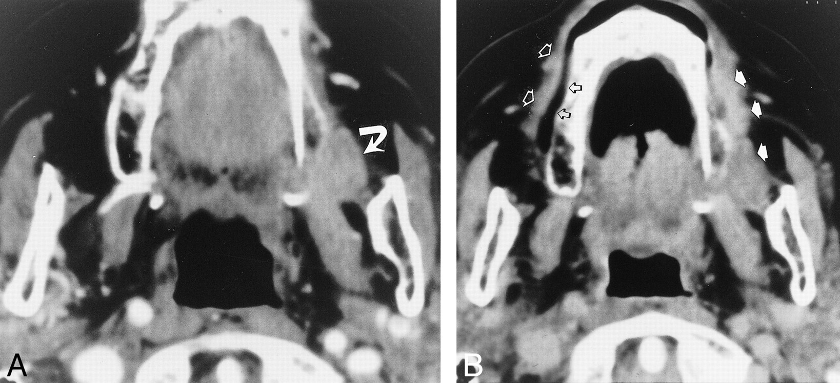
- fig 3.
64-year-old man with squamous cell carcinoma of the retromolar trigone. A, Conventional axial CT scan below the occlusal plane is normal. B, Puffed-cheek axial CT scan shows a small tumor in the left retromolar trigone (wavy arrow), inseparable from the mandible. The right retromolar trigone is normal (short thin arrow). The buccinator/buccal mucosa complex is thicker on the side of the tumor (short wide arrow) than on the normal (right) side
- fig 4.
77-year-old woman with squamous cell carcinoma of the maxilla. A, Conventional axial CT scan through the lower maxilla shows the tumor (arrow) inseparable from the gingival (alveolar) and buccal (cheek) mucosal surfaces. B, On the normal (right) side, the puffed-cheek axial CT scan separates the gingival mucosa and the alveolus (open black arrows) from the buccal mucosa and the buccinator muscle (open white arrows). The tumor thickens the cheek on the left side (solid arrows) and extends around the sulcus to the gingival mucosa. The combination of direct extension of tumor and decreased pliability of the cheek because of tumor invasion makes the two surfaces (buccal, gingival) inseparable
- fig 5.
86-year-old woman with verrucous hyperplasia of the buccal mucosa. A, Puffed-cheek axial CT scan (soft tissue algorithm) shows the exophytic mass (arrows). B, Puffed-cheek axial CT scan (bone algorithm) also shows the mass (short arrows). Only the bone algorithm shows the frenulum of the upper lip (long arrow), a normal structure. However, the soft tissue algorithm provides much greater detail of the tumor


In this issue


Jump to section
Related Articles
Cited By...
- Cross-Sectional Imaging of Third Molar-Related Abnormalities
- Imaging the oral cavity: key concepts for the radiologist
- Gauze Padding: A Simple Technique to Delineate Small Oral Cavity Tumors
- Evaluating "Eee" Phonation in Multidetector CT of the Neck
- Tumor Volume Assessment by 18F-FDG PET/CT in Patients with Oral Cavity Cancer with Dental Artifacts on CT or MR Images