Advertisement
  • Main menu
  • User menu
  • Search
  • English ▼
    • English
    • Afrikaans
    • Albanian
    • Amharic
    • Arabic
    • Armenian
    • Azerbaijani
    • Basque
    • Belarusian
    • Bengali
    • Bosnian
    • Bulgarian
    • Catalan
    • Cebuano
    • Chichewa
    • Chinese (Simplified)
    • Chinese (Traditional)
    • Corsican
    • Croatian
    • Czech
    • Danish
    • Dutch
    • Esperanto
    • Estonian
    • Filipino
    • Finnish
    • French
    • Frisian
    • Galician
    • Georgian
    • German
    • Greek
    • Gujarati
    • Haitian Creole
    • Hausa
    • Hawaiian
    • Hebrew
    • Hindi
    • Hmong
    • Hungarian
    • Icelandic
    • Igbo
    • Indonesian
    • Irish
    • Italian
    • Japanese
    • Javanese
    • Kannada
    • Kazakh
    • Khmer
    • Korean
    • Kurdish (Kurmanji)
    • Kyrgyz
    • Lao
    • Latin
    • Latvian
    • Lithuanian
    • Luxembourgish
    • Macedonian
    • Malagasy
    • Malay
    • Malayalam
    • Maltese
    • Maori
    • Marathi
    • Mongolian
    • Myanmar (Burmese)
    • Nepali
    • Norwegian
    • Pashto
    • Persian
    • Polish
    • Portuguese
    • Punjabi
    • Romanian
    • Russian
    • Samoan
    • Scottish Gaelic
    • Serbian
    • Sesotho
    • Shona
    • Sindhi
    • Sinhala
    • Slovak
    • Slovenian
    • Somali
    • Spanish
    • Sudanese
    • Swahili
    • Swedish
    • Tajik
    • Tamil
    • Telugu
    • Thai
    • Turkish
    • Ukrainian
    • Urdu
    • Uzbek
    • Vietnamese
    • Welsh
    • Xhosa
    • Yiddish
    • Yoruba
    • Zulu

Main menu

  • Home
  • Content
    • Current Issue
    • Accepted Manuscripts
    • Article Preview
    • Past Issue Archive
    • Video Articles
    • AJNR Case Collection
    • Case of the Week Archive
    • Case of the Month Archive
    • Classic Case Archive
  • Special Collections
    • Advancing NeuroMRI with High-Relaxivity Contrast Agents
    • AJNR Awards
    • Low-Field MRI
    • Alzheimer Disease
    • ASNR Foundation Special Collection
    • Photon-Counting CT
    • View All
  • Multimedia
    • AJNR Podcasts
    • AJNR SCANtastic
    • Trainee Corner
    • MRI Safety Corner
    • Imaging Protocols
  • For Authors
    • Submit a Manuscript
    • Submit a Video Article
    • Submit an eLetter to the Editor/Response
    • Manuscript Submission Guidelines
    • Statistical Tips
    • Fast Publishing of Accepted Manuscripts
    • Graphical Abstract Preparation
    • Imaging Protocol Submission
    • Author Policies
  • About Us
    • About AJNR
    • Editorial Board
    • Editorial Board Alumni
  • More
    • Become a Reviewer/Academy of Reviewers
    • Subscribers
    • Permissions
    • Alerts
    • Feedback
    • Advertisers
    • ASNR Home

User menu

  • Alerts
  • Log in

Search

  • Advanced search
American Journal of Neuroradiology

American Journal of Neuroradiology

ASHNR American Society of Functional Neuroradiology ASHNR American Society of Pediatric Neuroradiology ASSR
  • Alerts
  • Log in

Advanced Search

English ▼
  • English
  • Afrikaans
  • Albanian
  • Amharic
  • Arabic
  • Armenian
  • Azerbaijani
  • Basque
  • Belarusian
  • Bengali
  • Bosnian
  • Bulgarian
  • Catalan
  • Cebuano
  • Chichewa
  • Chinese (Simplified)
  • Chinese (Traditional)
  • Corsican
  • Croatian
  • Czech
  • Danish
  • Dutch
  • Esperanto
  • Estonian
  • Filipino
  • Finnish
  • French
  • Frisian
  • Galician
  • Georgian
  • German
  • Greek
  • Gujarati
  • Haitian Creole
  • Hausa
  • Hawaiian
  • Hebrew
  • Hindi
  • Hmong
  • Hungarian
  • Icelandic
  • Igbo
  • Indonesian
  • Irish
  • Italian
  • Japanese
  • Javanese
  • Kannada
  • Kazakh
  • Khmer
  • Korean
  • Kurdish (Kurmanji)
  • Kyrgyz
  • Lao
  • Latin
  • Latvian
  • Lithuanian
  • Luxembourgish
  • Macedonian
  • Malagasy
  • Malay
  • Malayalam
  • Maltese
  • Maori
  • Marathi
  • Mongolian
  • Myanmar (Burmese)
  • Nepali
  • Norwegian
  • Pashto
  • Persian
  • Polish
  • Portuguese
  • Punjabi
  • Romanian
  • Russian
  • Samoan
  • Scottish Gaelic
  • Serbian
  • Sesotho
  • Shona
  • Sindhi
  • Sinhala
  • Slovak
  • Slovenian
  • Somali
  • Spanish
  • Sudanese
  • Swahili
  • Swedish
  • Tajik
  • Tamil
  • Telugu
  • Thai
  • Turkish
  • Ukrainian
  • Urdu
  • Uzbek
  • Vietnamese
  • Welsh
  • Xhosa
  • Yiddish
  • Yoruba
  • Zulu
  • Home
  • Content
    • Current Issue
    • Accepted Manuscripts
    • Article Preview
    • Past Issue Archive
    • Video Articles
    • AJNR Case Collection
    • Case of the Week Archive
    • Case of the Month Archive
    • Classic Case Archive
  • Special Collections
    • Advancing NeuroMRI with High-Relaxivity Contrast Agents
    • AJNR Awards
    • Low-Field MRI
    • Alzheimer Disease
    • ASNR Foundation Special Collection
    • Photon-Counting CT
    • View All
  • Multimedia
    • AJNR Podcasts
    • AJNR SCANtastic
    • Trainee Corner
    • MRI Safety Corner
    • Imaging Protocols
  • For Authors
    • Submit a Manuscript
    • Submit a Video Article
    • Submit an eLetter to the Editor/Response
    • Manuscript Submission Guidelines
    • Statistical Tips
    • Fast Publishing of Accepted Manuscripts
    • Graphical Abstract Preparation
    • Imaging Protocol Submission
    • Author Policies
  • About Us
    • About AJNR
    • Editorial Board
    • Editorial Board Alumni
  • More
    • Become a Reviewer/Academy of Reviewers
    • Subscribers
    • Permissions
    • Alerts
    • Feedback
    • Advertisers
    • ASNR Home
  • Follow AJNR on Twitter
  • Visit AJNR on Facebook
  • Follow AJNR on Instagram
  • Join AJNR on LinkedIn
  • RSS Feeds

AJNR Awards, New Junior Editors, and more. Read the latest AJNR updates


Improved Turnaround Times | Median time to first decision: 12 days

Research ArticleBRAIN

Serial Proton MR Spectroscopic Imaging of Recurrent Malignant Gliomas after Gamma Knife Radiosurgery

Edward E. Graves, Sarah J. Nelson, Daniel B. Vigneron, Lynn Verhey, Michael McDermott, David Larson, Susan Chang, Michael D. Prados and William P. Dillon
American Journal of Neuroradiology April 2001, 22 (4) 613-624;
Edward E. Graves
  • Find this author on Google Scholar
  • Find this author on PubMed
  • Search for this author on this site
Sarah J. Nelson
  • Find this author on Google Scholar
  • Find this author on PubMed
  • Search for this author on this site
Daniel B. Vigneron
  • Find this author on Google Scholar
  • Find this author on PubMed
  • Search for this author on this site
Lynn Verhey
  • Find this author on Google Scholar
  • Find this author on PubMed
  • Search for this author on this site
Michael McDermott
  • Find this author on Google Scholar
  • Find this author on PubMed
  • Search for this author on this site
David Larson
  • Find this author on Google Scholar
  • Find this author on PubMed
  • Search for this author on this site
Susan Chang
  • Find this author on Google Scholar
  • Find this author on PubMed
  • Search for this author on this site
Michael D. Prados
  • Find this author on Google Scholar
  • Find this author on PubMed
  • Search for this author on this site
William P. Dillon
  • Find this author on Google Scholar
  • Find this author on PubMed
  • Search for this author on this site
  • Article
  • Figures & Data
  • Info & Metrics
  • Responses
  • References
  • PDF
Loading

Article Figures & Data

Figures

  • Tables
  • fig 1.
    • Download figure
    • Open in new tab
    • Download powerpoint
    fig 1.

    Patient 7.

    A–D, Serial contrast-enhanced gradient-echo images (2/6/1) and PRESS spectra (1000/144/1; voxel resolution, 1.0 cm3) acquired 1 day (A), 3 months (B), 5 months (C), and 6 months (D) after gamma knife treatment and aligned retrospectively. The 15-Gy isodose line is shown in red. The volume of contrast enhancement is significantly reduced at the 3-month time point, but increases at both the 5- and 6-month follow-ups. Reduction of Cho from baseline levels is apparent at the 3-, 5-, and 6-month time points. No significant change in NAA levels is observed, while a lactate/lipid resonance develops at 3 to 5 months but is reduced at 6 months. This patient received no further treatment and died 17 months after undergoing gamma knife radiosurgery.

  • fig 2.
    • Download figure
    • Open in new tab
    • Download powerpoint
    fig 2.

    Patient 11.

    A–C, Serial contrast-enhanced gradient echo-images (26/6/1) and PRESS spectra (1000/144/1; voxel resolution, 1.0 cm3) over the gamma knife target 3 days before (A) and 1 month (B) and 4 months (C) after radiosurgery. Two representative voxels are depicted, with the 15-Gy isodose line shown in red. The anterior voxel received an average radiation dose of 19.0 Gy, while the posterior voxel received 10.3 Gy. Reductions in both Cho and NAA as well as an increase in lactate/lipid are noted 1 month after radiosurgery. At 4 months, the less-irradiated voxel has developed a distinct tumorlike spectral pattern, while the heavily irradiated voxel appears necrotic. The lesion was resected, and brachytherapy seeds were implanted shortly after the last examination shown, but no biopsy samples were taken. The patient died 13 months after undergoing gamma knife treatment.
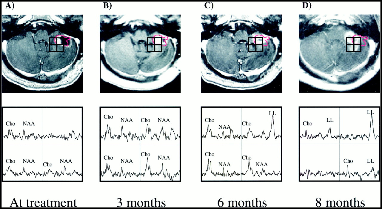
  • fig 3.
    • Download figure
    • Open in new tab
    • Download powerpoint
    fig 3.

    Patient 4.

    A–D, Serial contrast-enhanced gradient-echo images (26/6/1) and PRESS spectra (1000/144/1; voxel resolution, 1.0 cm3) of a cerebellar lesion 1 day before (A) and 3 months (B), 6 months (C), and 8 months (D) after treatment. The most irradiated voxel (almost completely within the red 15-Gy isodose line) received an average dose of 18.0 Gy, while the other three voxels received less than 10 Gy. At the time of radiosurgery low metabolite levels were noted in all voxels. Three months later, metabolite levels had increased, particularly within the radiation target. An increase in Cho is noted posterior to the radiation target at 3 and 6 months after treatment. At 8 months, these spectral abnormalities have become apparent radiologically, while the radiation target shows a necrotic spectrum. The development of a lactate/lipid peak within the high-dose region is also apparent from these serial observations. This patient was treated with gamma knife radiosurgery for a recurrence in the left superior frontal lobe 6 months after the initial treatment, and died 6 months thereafter.
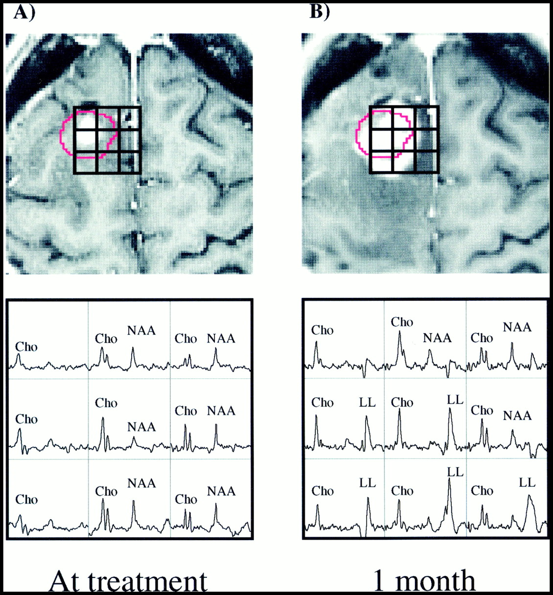
  • fig 4.
    • Download figure
    • Open in new tab
    • Download powerpoint
    fig 4.

    Patient 16.

    A and B, Surface-coil contrast-enhanced gradient-echo images (26/6/1) and PRESS spectra (1000/144/1; voxel resolution, 0.34 cm3) acquired 2 weeks before (A) and 1 month after (B) gamma knife radiosurgery and aligned retrospectively. Before radiosurgery, several voxels with Cho above that of normal-appearing tissue are present beyond the 15-Gy isodose line (shown in red) of the prescribed treatment plan. One month later, the Cho/NAA ratio in eight of the nine voxels shown has increased to greater than 1. Also conspicuous are the strong lactate/lipid resonances after treatment. One month after the last examination shown here, the patient underwent resection. Histologic analysis of biopsy samples taken during surgery showed residual/recurrent anaplastic astrocytoma.

  • fig 5.
    • Download figure
    • Open in new tab
    • Download powerpoint
    fig 5.

    Patient 18.

    A–C, Contrast-enhanced gradient-echo images (26/6/1) and PRESS spectra (1000/144/1; voxel resolution, 1.0 cm3) acquired 1 day before (A) and 2 months (B) and 4 months (C) after radiosurgery. Voxel shifting was used to obtain a spectrum centered within the 15-Gy isodose line (shown in red). Although a hemorrhage present at the time of gamma knife treatment complicated radiologic interpretation, elevated Cho was clearly well outside the gamma knife target. At the last measured time point, the Cho/NAA ratio of the treated voxel (average dose, 18.5 Gy) was reduced to values comparable to normal tissue, while tumorlike spectral patterns remained present posteriorly. Resection was recommended at this time, during which biopsy samples were obtained that verified the new contrast enhancement as residual/recurrent GBM, including some necrosis.
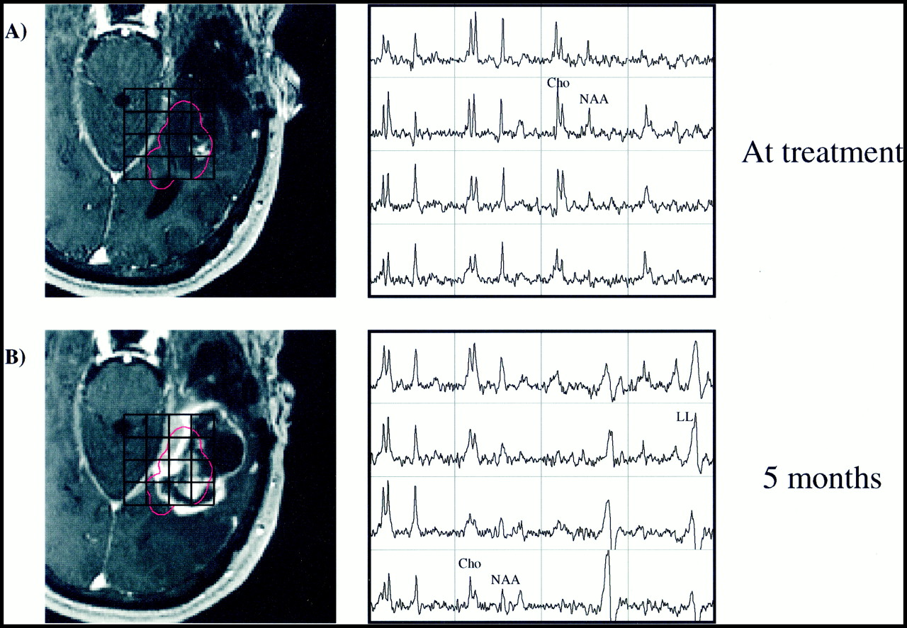
  • fig 6.
    • Download figure
    • Open in new tab
    • Download powerpoint
    fig 6.

    Patient 13.

    A and B, Contrast-enhanced gradient-echo images (26/6/1) and spectra (1000/144/1; voxel resolution, 1.0 cm3) acquired the day of radiosurgery (A) and 5 months afterward (B). The radiation target was prescribed to include spectral abnormalities beyond the regions of contrast enhancement. Despite the enlarged treatment field, the 6-month time point shows a large increase in contrast-enhancing volume. Increases in Cho within regions of new contrast enhancement suggest they correspond to recurrent tumor. Also conspicuous is the development of lactate/lipid resonances within the high-dose region. Resection was performed after the final time point shown, which histologically confirmed the increased contrast enhancement as recurrent tumor.

Tables

  • Figures
  • TABLE 1:
    • View popup
    • Download powerpoint
    TABLE 1:

    Patient data

  • TABLE 2:
    • View popup
    • Download powerpoint
    TABLE 2:

    Spectral response over the gamma knife target for the three response groups

  • TABLE 3:
    • View popup
    • Download powerpoint
    TABLE 3:

    Spectral evolution of regions of new contrast enhancement in patients with tumor recurrence

  • TABLE 4:
    • View popup
    • Download powerpoint
    TABLE 4:

    Biopsy results versus spectral measurements for six patients

PreviousNext
Back to top

In this issue

American Journal of Neuroradiology
Vol. 22, Issue 4
1 Apr 2001
  • Table of Contents
  • Index by author
Advertisement
Print
Download PDF
Email Article
Cite this article
0 Responses
Respond to this article
Share
Bookmark this article
Serial Proton MR Spectroscopic Imaging of Recurrent Malignant Gliomas after Gamma Knife Radiosurgery
Edward E. Graves, Sarah J. Nelson, Daniel B. Vigneron, Lynn Verhey, Michael McDermott, David Larson, Susan Chang, Michael D. Prados, William P. Dillon
American Journal of Neuroradiology Apr 2001, 22 (4) 613-624;
del.icio.us logo Twitter logo Facebook logo Mendeley logo
  • Tweet Widget

Jump to section

  • Article
    • Abstract
    • Methods
    • Results
    • Discussion
    • Conclusion
    • Acknowledgments
    • Footnotes
    • References
  • Figures & Data
  • Info & Metrics
  • Responses
  • References
  • PDF

Related Articles

  • No related articles found.
  • PubMed
  • Google Scholar

Cited By...

  • Early Metabolic Changes in 1H-MRSI Predictive for Survival in Patients With Newly Diagnosed High-grade Glioma
  • Role of MRI in Primary Brain Tumor Evaluation
  • Specific biomarkers of receptors, pathways of inhibition and targeted therapies: clinical applications
  • Changes in Serial Magnetic Resonance Spectroscopy Predict Outcome in High-grade Glioma During and After Postoperative Radiotherapy
  • Permeability Estimates in Histopathology-Proved Treatment-Induced Necrosis Using Perfusion CT: Can These Add to Other Perfusion Parameters in Differentiating from Recurrent/Progressive Tumors?
  • Distinguishing Recurrent Intra-Axial Metastatic Tumor from Radiation Necrosis Following Gamma Knife Radiosurgery Using Dynamic Susceptibility-Weighted Contrast-Enhanced Perfusion MR Imaging
  • The Extent and Severity of Vascular Leakage as Evidence of Tumor Aggressiveness in High-Grade Gliomas
  • Diagnosis and Treatment of Recurrent High-Grade Astrocytoma
  • Physiologic and Metabolic Magnetic Resonance Imaging in Gliomas
  • NMR Spectroscopy and Pediatric Brain Tumors
  • Crossref
  • Google Scholar

This article has not yet been cited by articles in journals that are participating in Crossref Cited-by Linking.

More in this TOC Section

  • Multimodal CT Provides Improved Performance for Lacunar Infarct Detection
  • Optimal MRI Sequence for Identifying Occlusion Location in Acute Stroke: Which Value of Time-Resolved Contrast-Enhanced MRA?
  • Evaluating the Effects of White Matter Multiple Sclerosis Lesions on the Volume Estimation of 6 Brain Tissue Segmentation Methods
Show more Brain

Similar Articles

Advertisement

Indexed Content

  • Current Issue
  • Accepted Manuscripts
  • Article Preview
  • Past Issues
  • Editorials
  • Editor's Choice
  • Fellows' Journal Club
  • Letters to the Editor
  • Video Articles

Cases

  • Case Collection
  • Archive - Case of the Week
  • Archive - Case of the Month
  • Archive - Classic Case

More from AJNR

  • Trainee Corner
  • Imaging Protocols
  • MRI Safety Corner
  • Book Reviews

Multimedia

  • AJNR Podcasts
  • AJNR Scantastics

Resources

  • Turnaround Time
  • Submit a Manuscript
  • Submit a Video Article
  • Submit an eLetter to the Editor/Response
  • Manuscript Submission Guidelines
  • Statistical Tips
  • Fast Publishing of Accepted Manuscripts
  • Graphical Abstract Preparation
  • Imaging Protocol Submission
  • Evidence-Based Medicine Level Guide
  • Publishing Checklists
  • Author Policies
  • Become a Reviewer/Academy of Reviewers
  • News and Updates

About Us

  • About AJNR
  • Editorial Board
  • Editorial Board Alumni
  • Alerts
  • Permissions
  • Not an AJNR Subscriber? Join Now
  • Advertise with Us
  • Librarian Resources
  • Feedback
  • Terms and Conditions
  • AJNR Editorial Board Alumni

American Society of Neuroradiology

  • Not an ASNR Member? Join Now

© 2025 by the American Society of Neuroradiology All rights, including for text and data mining, AI training, and similar technologies, are reserved.
Print ISSN: 0195-6108 Online ISSN: 1936-959X

Powered by HighWire
Email this Article

Thank you for your interest in spreading the word on American Journal of Neuroradiology.

NOTE: We only request your email address so that the person you are recommending the page to knows that you wanted them to see it, and that it is not junk mail. We do not capture any email address.

Enter multiple addresses on separate lines or separate them with commas.
Serial Proton MR Spectroscopic Imaging of Recurrent Malignant Gliomas after Gamma Knife Radiosurgery
(Your Name) has sent you a message from American Journal of Neuroradiology
(Your Name) thought you would like to see the American Journal of Neuroradiology web site.
CAPTCHA
This question is for testing whether or not you are a human visitor and to prevent automated spam submissions.
Cite this article
Edward E. Graves, Sarah J. Nelson, Daniel B. Vigneron, Lynn Verhey, Michael McDermott, David Larson, Susan Chang, Michael D. Prados, William P. Dillon
Serial Proton MR Spectroscopic Imaging of Recurrent Malignant Gliomas after Gamma Knife Radiosurgery
American Journal of Neuroradiology Apr 2001, 22 (4) 613-624;

Citation Manager Formats

  • BibTeX
  • Bookends
  • EasyBib
  • EndNote (tagged)
  • EndNote 8 (xml)
  • Medlars
  • Mendeley
  • Papers
  • RefWorks Tagged
  • Ref Manager
  • RIS
  • Zotero

We use cookies on this site to enhance your user experience. By clicking any link on this page you are giving your consent for us to set cookies.