
Article Figures & Data
Figures
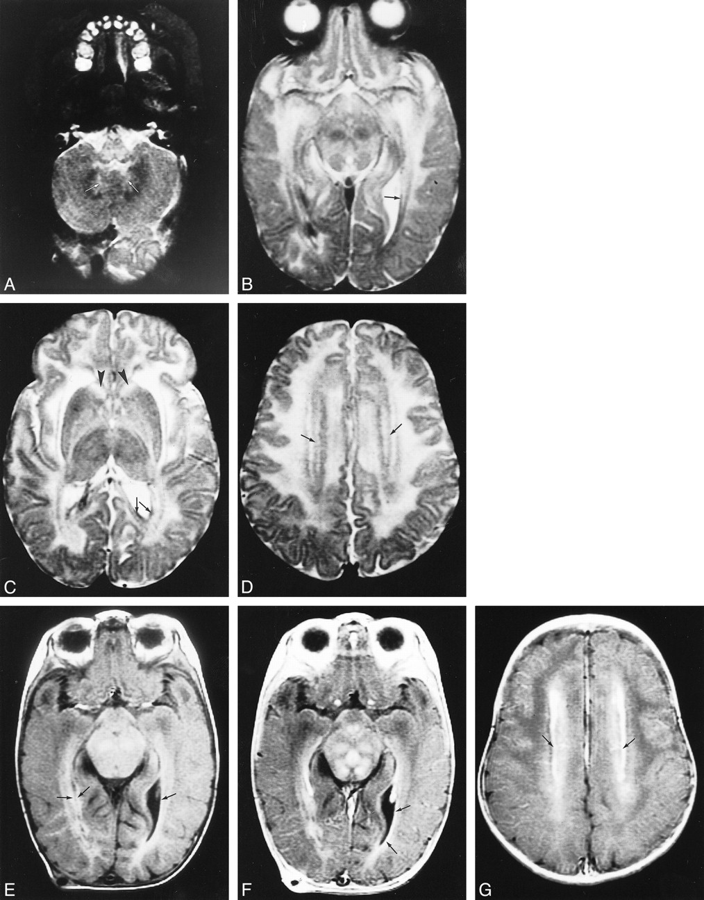
- fig 1.
Early MR imaging study at the age of 4 months in a patient with autopsy-proved infantile Alexander disease.
A–D, T2-weighted images show abnormally high signal in the medulla (A), the hilus of the dentate nucleus (arrows, A), the entire midbrain except for the red nuclei (B), the basal ganglia, and the thalamus (C). The frontal white matter has a slightly higher signal intensity than the occipital white matter (C). The head of the caudate nucleus is swollen (arrowheads, C). Around the ventricles, there is a rim of low signal intensity (arrows, B–D).
E–G, T1-weighted images show high signal intensity of the periventricular rim (arrows, E). After contrast administration, the T1-weighted images show enhancement of areas in the midbrain (F), ventricular lining (arrows, F), and periventricular rim (arrows, G).
- fig 2.
A–D, Early (A) and late (B–D) MR studies of a patient with autopsy-confirmed juvenile Alexander disease, obtained at ages 20 months (A) and 9 years (B–D). The early T2-weighted image (A) shows extensive cerebral white matter abnormalities with partial sparing of the occipital region. There is a thin periventricular rim of low signal intensity (arrows, A). The basal ganglia and thalamus have an increased signal intensity. The putamen and caudate nucleus are mildly swollen (A). On follow-up, the extent of the cerebral white matter abnormalities is more or less the same; the occipital white matter is still partially spared (D). The basal nuclei are dark and atrophic on the T2-weighted images (D). A thin periventricular rim of low signal intensity is visible (arrows, D). The proton density–weighted image (C) shows enormous cysts in the frontoparietal white matter, a large cavum vergae, and enlarged lateral ventricles. A lesion is seen in the posterior part of the medulla (B)
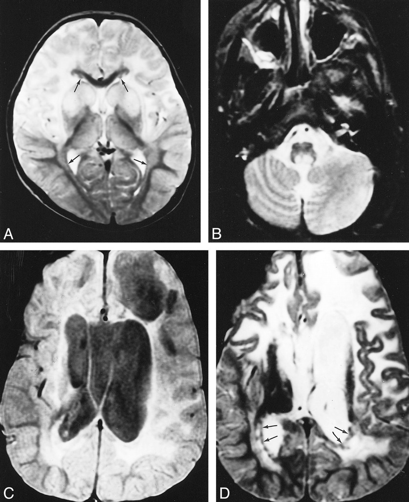
- fig 3.
MR imaging of a patient with biopsy-confirmed infantile Alexander disease.
A and B, At the age of 1½ months, the frontal white matter has a slightly higher signal intensity on T2-weighted images and slightly lower signal intensity on unenhanced T1-weighted images than does the remainder of the cerebral white matter, which has normal signal intensity for unmyelinated white matter. There is a periventricular rim of low signal intensity on T2-weighted images (arrows, A) and high signal intensity on T1-weighted images (arrows, B), with some extensions into the frontal white matter (arrowheads, A and B). The caudate nucleus and putamen have high signal intensity on T2-weighted images and are mildly swollen.
C and D, At the age of 3 months, a major increase in ventricular size is seen with extreme thinning of the posterior cerebral mantle. The frontal white matter has a more abnormal signal intensity than the occipital white matter, appears markedly swollen, and shows early cystic degeneration (arrows, D). There is a thin periventricular rim of low signal intensity on T2-weighted images (arrows, C). The basal ganglia are now markedly atrophic. After contrast administration, enhancement occurs in the ventricular lining, caudate nucleus, putamen, frontal white matter, and parts of the frontal cortex (D).
- fig 4.
Early MR imaging studies in a patient with presumed juvenile Alexander disease, obtained at the age of 4 years.
A and B, Extensive cerebral white matter abnormalities are seen on these T2-weighted images (B), with sparing of the occipital U fibers (arrows, B). The signal abnormality is more pronounced in the frontal than in the occipital white matter. There is an irregular periventricular rim of low signal intensity (arrowheads, B). The basal ganglia and thalamus have a mildly increased signal intensity. Within the posterior fossa, signal abnormalities are seen in the central part of the medulla, the hilus of the dentate nucleus, and the cerebellar hemispheric white matter, characteristically with the normal dentate nucleus prominently visible in between (A).
fig 5. Contrast-enhanced MR image in a patient with presumed juvenile Alexander disease, obtained at the age of 12 years. Note enhancement of the intraparenchymal trajectory of the fifth cranial nerve on both sides
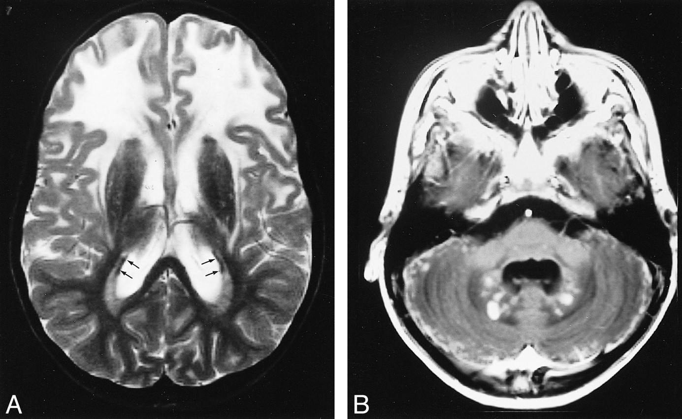
- fig 6.
Late MR imaging study of a patient with presumed juvenile Alexander disease, obtained at the age of 10 years.
A and B, There is extensive white matter involvement with frontal preponderance (A). The basal ganglia are dark and atrophic on T2-weighted images (A). A thin periventricular rim of low signal intensity is just visible (arrows, A). After contrast administration, enhancement of the entire cerebellar surface and dentate nucleus is seen (B).


Tables
 TABLE 1:
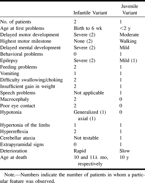
Clinical signs and symptoms in patients with a histo~logically confirmed diagnosis
- TABLE 2:
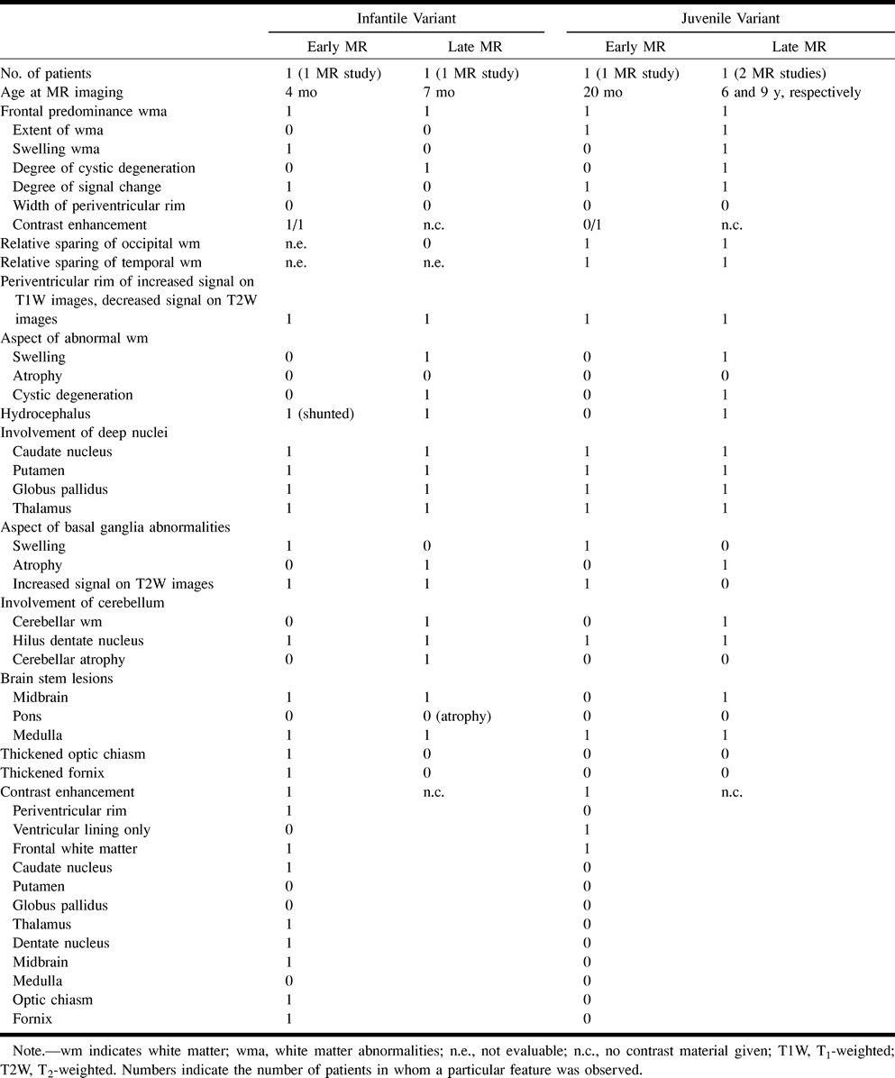
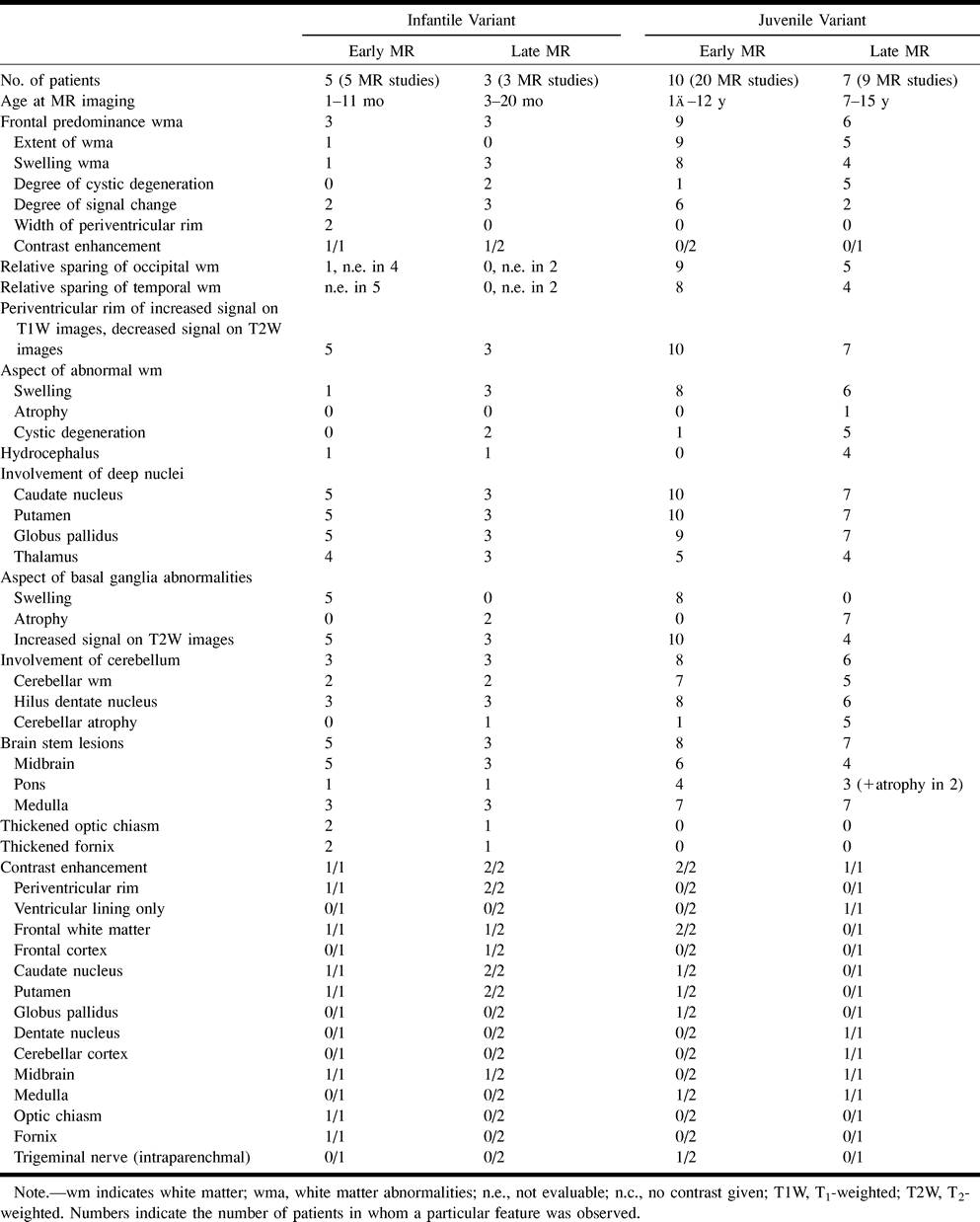
MR findings in patients with a histologically confirmed diagnosis
- TABLE 3:
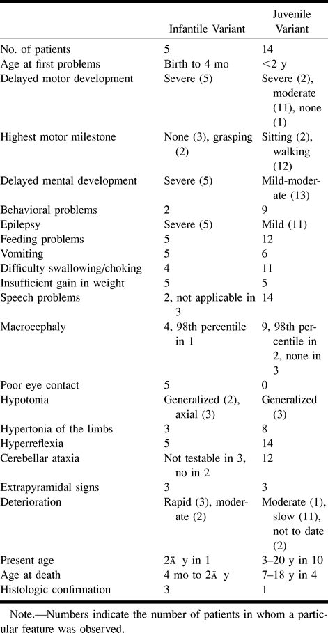
Clinical signs and symptoms in patients with an MR imaging-based diagnosis
In this issue


Jump to section
Related Articles
Cited By...
- Diagnosing Alexander disease in adults
- Gfap Mutation and Astrocyte Dysfunction Lead to a Neurodegenerative Profile with Impaired Synaptic Plasticity and Cognitive Deficits in a Rat Model of Alexander Disease
- Brainstem Chipmunk Sign: A Diagnostic Imaging Clue across All Subtypes of Alexander Disease
- Adult-Onset Alexander Disease: New Causal Sequence Variant in the GFAP Gene
- Teaching NeuroImage: Dorsal Medullary Lesions in Juvenile-Onset Alexander Disease
- Pearls & Oy-sters: Adult-Onset Alexander Disease With Transient Swelling of the Medulla Oblongata
- Antisense therapy in a new rat model of Alexander disease reverses GFAP pathology, white matter deficits, and motor impairment
- A novel mutation in the GFAP gene expands the phenotype of Alexander disease
- Structural basis for the inhibition of translation through eIF2{alpha} phosphorylation
- Brain MR Imaging Findings in Woodhouse-Sakati Syndrome
- CSF and Blood Levels of GFAP in Alexander Disease
- Megalencephalic leucoencephalopathy with subcortical cysts: subcortical diffuse leucoencephalopathy associated with white matter cystic degeneration
- A practical approach to diagnosing adult onset leukodystrophies
- Neuroimaging and clinical features in type II (late-onset) Alexander disease
- Alexander Disease
- GFAP mutations, age at onset, and clinical subtypes in Alexander disease
- Invited Article: An MRI-based approach to the diagnosis of white matter disorders
- Alexander disease: Not just a leukodystrophy anymore
- Alexander disease: Ventricular garlands and abnormalities of the medulla and spinal cord
- The clinicopathological spectrum of Rosenthal fibre encephalopathy and Alexander's disease: a case report and review of the literature
- Molecular findings in symptomatic and pre-symptomatic Alexander disease patients