
Article Figures & Data
Figures
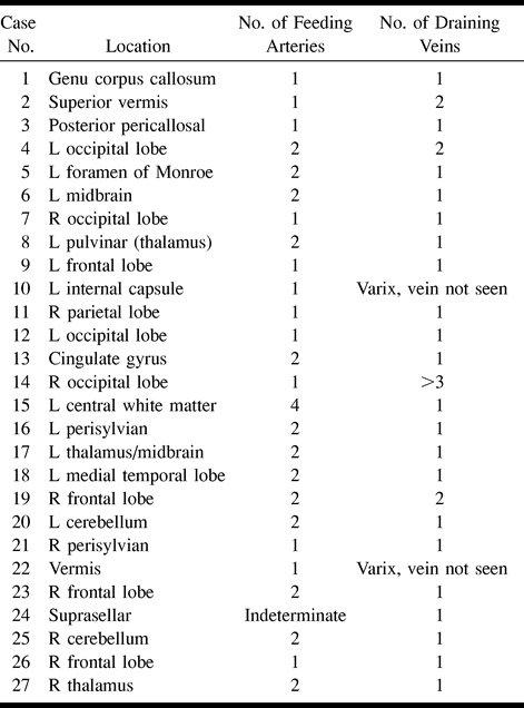
- fig 1.
Case 14.
A and B, Vertebral angiograms (lateral projections). The first angiogram (A), 3 months after an intracerebral hemorrhage, shows a 2- to 3-cm right occipital AVM. The second angiogram (B), after a further 3 months, at the time of admission for STRS, shows absence of the previously visible AVM.
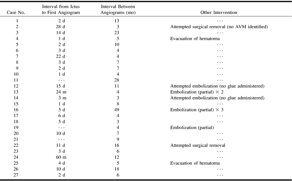
- fig 2.
Case 15.
A and B, Left internal carotid angiograms (frontal projections) show absence of the AVM after an interval of 8 months. The patient presented with hemiplegia (Glasgow Coma Scale score = 4) associated with an intracerebral hematoma.
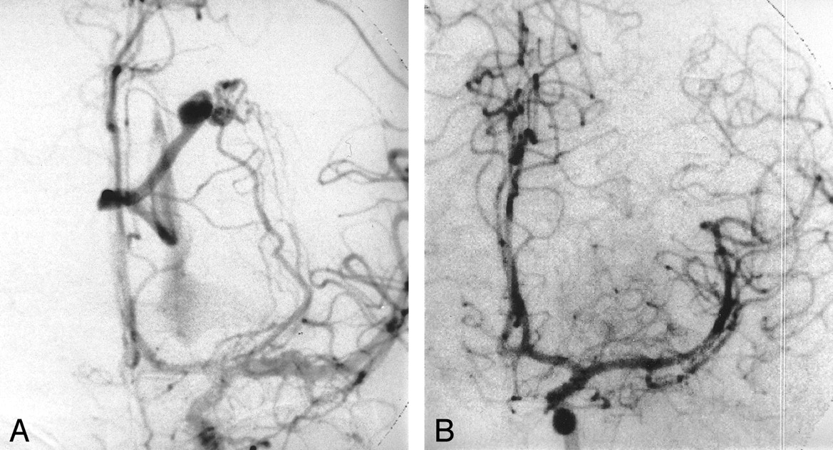
- fig 3.
Case 6.
A–C, Vertebral angiograms (lateral projections) at presentation (A), at presentation for STRS 4 months later (B), and at time of recurrence 6 years later (C). A barely visible AVM is shown with early shunting of contrast medium into the straight sinus (A). No AVM is seen 4 months later (B). Six years later, the angiogram shows an AVM in the same location but with predominate early filling of the superior vermian vein (C).


Tables
 TABLE 1:
Location and number of vessels associated with spon~taneously obliterated AVMs
- TABLE 2:
Presentation, morphologic characteristics, and follow-up data for patients with spontaneously obliterated AVMs
- TABLE 3:
Intervention and timing of obliteration associated with spontaneously obliterated AVMs


