Advertisement

Main menu

  • Home
  • Content
    • Current Issue
    • Accepted Manuscripts
    • Article Preview
    • Past Issue Archive
    • Video Articles
    • AJNR Case Collection
    • Case of the Week Archive
    • Case of the Month Archive
    • Classic Case Archive
  • Special Collections
    • AJNR Awards
    • Low-Field MRI
    • Alzheimer Disease
    • ASNR Foundation Special Collection
    • Photon-Counting CT
    • View All
  • Multimedia
    • AJNR Podcasts
    • AJNR SCANtastic
    • Trainee Corner
    • MRI Safety Corner
    • Imaging Protocols
  • For Authors
    • Submit a Manuscript
    • Submit a Video Article
    • Submit an eLetter to the Editor/Response
    • Manuscript Submission Guidelines
    • Statistical Tips
    • Fast Publishing of Accepted Manuscripts
    • Graphical Abstract Preparation
    • Imaging Protocol Submission
    • Author Policies
  • About Us
    • About AJNR
    • Editorial Board
    • Editorial Board Alumni
  • More
    • Become a Reviewer/Academy of Reviewers
    • Subscribers
    • Permissions
    • Alerts
    • Feedback
    • Advertisers
    • ASNR Home

User menu

  • Alerts
  • Log in

Search

  • Advanced search
American Journal of Neuroradiology

American Journal of Neuroradiology

ASHNR American Society of Functional Neuroradiology ASHNR American Society of Pediatric Neuroradiology ASSR
  • Alerts
  • Log in

Advanced Search

  • Home
  • Content
    • Current Issue
    • Accepted Manuscripts
    • Article Preview
    • Past Issue Archive
    • Video Articles
    • AJNR Case Collection
    • Case of the Week Archive
    • Case of the Month Archive
    • Classic Case Archive
  • Special Collections
    • AJNR Awards
    • Low-Field MRI
    • Alzheimer Disease
    • ASNR Foundation Special Collection
    • Photon-Counting CT
    • View All
  • Multimedia
    • AJNR Podcasts
    • AJNR SCANtastic
    • Trainee Corner
    • MRI Safety Corner
    • Imaging Protocols
  • For Authors
    • Submit a Manuscript
    • Submit a Video Article
    • Submit an eLetter to the Editor/Response
    • Manuscript Submission Guidelines
    • Statistical Tips
    • Fast Publishing of Accepted Manuscripts
    • Graphical Abstract Preparation
    • Imaging Protocol Submission
    • Author Policies
  • About Us
    • About AJNR
    • Editorial Board
    • Editorial Board Alumni
  • More
    • Become a Reviewer/Academy of Reviewers
    • Subscribers
    • Permissions
    • Alerts
    • Feedback
    • Advertisers
    • ASNR Home
  • Follow AJNR on Twitter
  • Visit AJNR on Facebook
  • Follow AJNR on Instagram
  • Join AJNR on LinkedIn
  • RSS Feeds

AJNR Awards, New Junior Editors, and more. Read the latest AJNR updates

Research ArticleHead and Neck Imaging

Parapharyngeal Second Branchial Cyst Manifesting as Cranial Nerve Palsies: MR Findings

Ji Hoon Shin, Ho Kyu Lee, Sang Yoon Kim, Hyung Wook Park, Shin Kwang Khang, Choon Gon Choi and Dae Chul Suh
American Journal of Neuroradiology March 2001, 22 (3) 510-512;
Ji Hoon Shin
  • Find this author on Google Scholar
  • Find this author on PubMed
  • Search for this author on this site
Ho Kyu Lee
  • Find this author on Google Scholar
  • Find this author on PubMed
  • Search for this author on this site
Sang Yoon Kim
  • Find this author on Google Scholar
  • Find this author on PubMed
  • Search for this author on this site
Hyung Wook Park
  • Find this author on Google Scholar
  • Find this author on PubMed
  • Search for this author on this site
Shin Kwang Khang
  • Find this author on Google Scholar
  • Find this author on PubMed
  • Search for this author on this site
Choon Gon Choi
  • Find this author on Google Scholar
  • Find this author on PubMed
  • Search for this author on this site
Dae Chul Suh
  • Find this author on Google Scholar
  • Find this author on PubMed
  • Search for this author on this site
  • Article
  • Figures & Data
  • Info & Metrics
  • Responses
  • References
  • PDF
Loading

Abstract

Summary: We report the MR findings of parapharyngeal branchial cleft cyst manifesting as multiple, lower cranial nerve palsies in a 35-year-old woman. On MR images, a well-marginated cystic mass was detected in the right parapharyngeal space, with displacement of both the right internal carotid artery and the right internal jugular vein on the posterolateral side. The cyst contained a whitish fluid that was slightly hyperintense on T1-weighted images and slightly hypointense to CSF on T2-weighted images. No enhancement on contrast-enhanced T1-weighted images was present. The right side of the tongue showed high signal intensity on T2-weighted images, suggesting denervation.

Branchial cleft cysts are the most common neck masses in adults. Most are second branchial cleft cysts, which occur in the neck, anterior to the sternocleidomastoid muscle at the mandibular angle. Rarely these cysts may be present in the parapharyngeal space. We describe a case of parapharyngeal branchial cleft cyst manifesting as multiple lower cranial nerve palsies, an unusual location and presentation for this lesion.

Case Report

A 35-year-old woman initially presented with a 1-month history of hoarseness. On physical examination, the gag reflex was decreased, the uvula was deviated to the left, and a right vocal cord palsy and right-sided paresis of the tongue were noted and attributed to palsies of cranial nerves IX, X, and XII. There was no palpable abnormality of the neck, and the patient was afebrile.

MR scanning revealed a large, thin-walled cyst in the right parapharyngeal space, the content of which was slightly hyperintense on T1-weighted images and slightly hypointense to CSF on T2-weighted images (Fig 1A and B). There was no enhancement of the lesion on contrast-enhanced T1-weighted images (Fig 1C). The cyst displaced both the right internal carotid artery and the right internal jugular vein on the posterolateral side. The cyst occupied the right parapharyngeal space from below the skull base to the hyoid bone level on coronal images (Fig 1D). The right side of the tongue showed high signal intensity on short inversion time inversion-recovery (STIR) images (Fig 1E), consistent with denervation. There were no abnormal lymph nodes in either neck area.

fig 1.
  • Download figure
  • Open in new tab
  • Download powerpoint
fig 1.

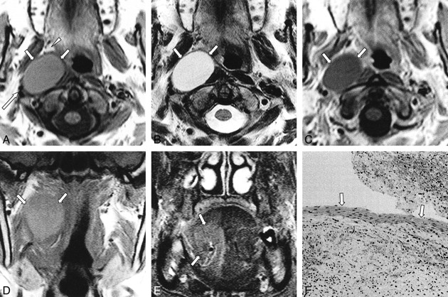
A 35-year-old woman with hoarseness.

A, T1-weighted (627/15 [TR/TE]) axial MR image shows well-marginated mass (arrows) slightly hyperintense to the CSF in the right parapharyngeal space. The mass displaces parapharyngeal fat anteriorly (arrowhead) and compresses the lateral portion of the longus colli muscle. The mass also displaces both the right internal carotid artery and the right internal jugular vein to its posterolateral side (long arrow).

B, T2-weighted (3800/99) axial MR image shows the mass (arrows) is slightly hypointense to the CSF.

C, Contrast-enhanced T1-weighted (627/15) axial MR image reveals no definite enhancement of the mass (arrows).

D, T1-weighted (530/12) coronal MR image shows the mass (arrows) occupies the right parapharyngeal space from below the skull base to the hyoid bone level.

E, The STIR (6500/150/60 [TR/TI/TE]) coronal image shows the right side of the tongue has high signal intensity (arrows), suggesting denervation.

F, Photomicrograph (hematoxylin-eosin, original magnification ×100) of the lesion shows a squamous epithelial-lined cyst wall (arrows), consistent with a branchial cleft cyst.

Excision of the neck mass was performed via a transcervical approach. A 5-cm × 4-cm, well-circumscribed, whitish ovoid mass was identified in the right parapharyngeal space. The mass was cystic and contained thick whitish pus. The adjacent cranial nerves IX, X, and XII were compressed by the mass, but were grossly normal in size and color. Histopathologic evaluation of the mass revealed a squamous epithelial-lined cyst with lymphoid infiltrate, consistent with a branchial cleft cyst (Fig 1F). The mass was identified as an infected second branchial cleft cyst. Gradual recovery of the multiple cranial nerve palsies occurred within 3 weeks.

Discussion

Second branchial cleft remnants account for the majority of branchial cleft abnormalities. Embryologically, the second arch overgrows the second, third, and fourth branchial clefts. This process results in expansion of the second branchial cleft into an elongated common cavity, called the cervical sinus of His, which is obliterated shortly after its formation. Various degrees of incomplete closure of the sinus lead to anomalies of the second branchial cleft. Anomalies can occur anywhere along an embryologically defined tract that extends from the external opening, the anterior border of the junction of the middle and lower thirds of the sternocleidomastoid muscle, passes between the internal and external carotid arteries superficial to cranial nerves IX and XII, and enters the oropharyngeal tonsillar fossa (1, 2). The parapharyngeal space is a very rare location for a branchial cleft cyst. This rare location superior to the tonsillar fossa can best be explained by the fact that a second branchial cleft cyst can have an associated tract that passes to the palatine tonsil. Because this tract passes through the caudal paraphyrngeal space, cysts that occur in this location are usually classified as second cleft anomalies (3). Although two previous reports stated that his type of parapharyngeal cyst was thought to be a first branchial cleft anomaly (4, 5), the site of pharyngeal attachment and lack of deformity of derivatives of the first branchial apparatus support the view that such cysts originate in the second branchial apparatus, as in our study (6).

Symptoms referable to masses within the parapharyngeal space are often minimal; the most common is a bulge in the overlying pharynx (2). Palsy of cranial nerves IX, X, XI, or XII is extremely rare in benign tumors or cysts, and usually suggests direct infiltration of local malignancies or metastatic lymph nodes (7). In our patient, the paralysis of lower cranial nerves IX (decreased gag reflex), X (uvula deviation and hoarseness secondary to vocal cord palsy), and XII (tongue deviation) can be related to mechanical compression of nerves as a result of marked enlargement of the cyst attributable to superimposed infection (8). At surgery, the cranial nerves showed grossly normal morphologic characteristics and color, although they were compressed by the mass. The cranial nerve palsies subsided in a relatively short period probably because the nerves manifested a transitory dysfunction owing to mechanical compression. There have been few reports on cranial nerve palsies associated with branchial cleft cysts (2, 8)

The reported MR findings of branchial cleft cysts do not differ from those of other cysts; ie, a well-marginated, homogeneous, cystic mass. The signal intensity is isointense to the CSF on T1- and T2-weighted images (3, 6). If the cyst is infected, a thin rim of enhancement is possible. Although this was not observed in our case, the margin of the cyst could also be poorly circumscribed because the adjacent fat planes may be obscured, and the signal intensity of the cystic content may be increased on T1-weighted images because of the increased protein content of the cystic fluid (2). An important goal of imaging is to differentiate primary parapharyngeal space lesions from more common, deep-lobe parotid lesions that have infiltrated the space in order to determine the appropriate surgical approach (9). MR imaging can accomplish this by revealing the fat plane between the normal parotid gland and a mass arising within the parapharyngeal space.

The differential diagnosis of a parapharyngeal mass includes cystic schwannomas and minor salivary gland tumors. Although large schwannomas can undergo cystic degeneration, complete cystic change preserving an even, thin wall is very rare, and good enhancement of the solid portion is seen in most cases. Furthermore, anteromedial displacement of the internal carotid artery is frequently present with schwannomas in this region and is highly suggestive of an origin within the carotid space (10). Minor salivary gland tumors, such as pleomorphic adenomas originating within the prestyloid parapharyngeal space, can be distinguished from a branchial cleft cyst because they usually show heterogeneous, intermediate signal intensity on T1-weighted images and intermediate-to-high signal intensity on T2-weighted images, even though they also posteriorly displace the internal carotid artery (9, 10). Furthermore, complete cystic change is very rare in minor salivary gland tumors.

Complete surgical excision is the treatment of choice for preventing recurrence (11), although percutaneous ethanol sclerotherapy could also be used (12). The transcervical approach provides the most complete exposure (11).

In conclusion, the differential diagnosis of a cystic lesion within the parapharyngeal space should include an atypical second branchial cleft cyst. A rapidly growing, infected branchial cleft cyst can present with lower cranial nerve palsies.

Footnotes

  • 1 Address reprint requests to Ho Kyu Lee, M.D., Department of Radiology, Asan Medical Center, 388-1 Poongnap-Dong, Songpa-Ku, Seoul 138-736, South Korea.

References

  1. ↵
    Papay FA, Kalucis C, Eliachar I, Tucker HM. Nasopharyngeal presentation of second branchial cleft cyst. Otolaryngol Head Neck Surg 1994;110:232-234
    CrossRefPubMed
  2. ↵
    Durrant TJ, Sevick RJ, Lauryssen C, MacRae ME. Parapharyngeal branchial cleft cyst presenting with cranial nerve palsies. Can Assoc Radiol J 1994;45:134-136
    PubMed
  3. ↵
    Som PM, Brandwein MS, Silvers A. Nodal inclusion cysts of the parotid gland and parapharyngeal space: a discussion of lymphoepithelial, AIDS-related parotid, and branchial cysts, cystic Warthin's tumors, and cysts in Sjogren's syndrome. Laryngoscope 1995;105:1122-1128
    PubMed
  4. ↵
    Guissani M. Cisti rinofaringea di origine branchiale. Annali di Laringologia, Otologia, Rinologia, Faringologia 1928;29:213
  5. Hoogland GA. Nasopharyngeal cyst of branchiogenic origin. J Laryngol Otol 1951;65:515-517
  6. ↵
    Shidara K, Uruma T, Yasuoka Y, Kamei T. Two cases of nasopharyngeal branchial cyst. J Laryngol Otol 1993;107:453-455
    PubMed
  7. ↵
    Sobol SM. Benign tumors. In: Thawley SE, Panje WR, eds. Comprehensive Management of Head and Neck Tumors. Philadelphia: WB Saunders 1987;1353-1383
  8. ↵
    Gatot A, Tovi F, Fliss DM, Tanai-Inbar I. Branchial cleft cyst manifesting as hypoglossal nerve palsy. Head Neck 1991;13:249-250
    PubMed
  9. ↵
    Som PM, Sacher M, Stollman AL, Biller HF, Lawson W. Common tumors of the parapharyngeal space: refined imaging diagnosis. Radiology 1988;169:81-85
    PubMed
  10. ↵
    Tom BM, Rao VM, Guglielmo F. Imaging of the parapharyngeal space: anatomy and pathology. Crit Rev Diagn Imaging 1991;31:315-356
    PubMed
  11. ↵
    Guneri A, Gunbay MU, Guneri EA, Ceryan K, Sutay S. Management of parapharyngeal space cysts. J Laryngol Otol 1994;108:795-797
    PubMed
  12. ↵
    Fukumoto K, Kojima T, Tomonari H, Kontani K, Murai S, Tsujimoto F. Ethanol injection sclerotherapy for Baker's cyst, thyroglossal duct cyst, and branchial cleft cyst. Ann Plast Surg 1994;33:615-619
    CrossRefPubMed
  • Received August 2, 2000.
  • Copyright © American Society of Neuroradiology
View Abstract
PreviousNext
Back to top

In this issue

American Journal of Neuroradiology
Vol. 22, Issue 3
1 Mar 2001
  • Table of Contents
  • Index by author
Advertisement
Print
Download PDF
Email Article

Thank you for your interest in spreading the word on American Journal of Neuroradiology.

NOTE: We only request your email address so that the person you are recommending the page to knows that you wanted them to see it, and that it is not junk mail. We do not capture any email address.

Enter multiple addresses on separate lines or separate them with commas.
Parapharyngeal Second Branchial Cyst Manifesting as Cranial Nerve Palsies: MR Findings
(Your Name) has sent you a message from American Journal of Neuroradiology
(Your Name) thought you would like to see the American Journal of Neuroradiology web site.
CAPTCHA
This question is for testing whether or not you are a human visitor and to prevent automated spam submissions.
Cite this article
Ji Hoon Shin, Ho Kyu Lee, Sang Yoon Kim, Hyung Wook Park, Shin Kwang Khang, Choon Gon Choi, Dae Chul Suh
Parapharyngeal Second Branchial Cyst Manifesting as Cranial Nerve Palsies: MR Findings
American Journal of Neuroradiology Mar 2001, 22 (3) 510-512;

Citation Manager Formats

  • BibTeX
  • Bookends
  • EasyBib
  • EndNote (tagged)
  • EndNote 8 (xml)
  • Medlars
  • Mendeley
  • Papers
  • RefWorks Tagged
  • Ref Manager
  • RIS
  • Zotero
0 Responses
Respond to this article
Share
Bookmark this article
Parapharyngeal Second Branchial Cyst Manifesting as Cranial Nerve Palsies: MR Findings
Ji Hoon Shin, Ho Kyu Lee, Sang Yoon Kim, Hyung Wook Park, Shin Kwang Khang, Choon Gon Choi, Dae Chul Suh
American Journal of Neuroradiology Mar 2001, 22 (3) 510-512;
del.icio.us logo Twitter logo Facebook logo Mendeley logo
  • Tweet Widget
  • Facebook Like
  • Google Plus One

Jump to section

  • Article
    • Abstract
    • Case Report
    • Discussion
    • Footnotes
    • References
  • Figures & Data
  • Info & Metrics
  • Responses
  • References
  • PDF

Related Articles

  • No related articles found.
  • PubMed
  • Google Scholar

Cited By...

  • No citing articles found.
  • Crossref
  • Google Scholar

This article has not yet been cited by articles in journals that are participating in Crossref Cited-by Linking.

More in this TOC Section

  • FDG Uptake and Thyroid Cancer Post-BRAF Therapy
  • Normal Facial Nerve Enhancement on VIBE Sequence
  • Lipochoristomas and inner ear signal
Show more Head and Neck Imaging

Similar Articles

Advertisement

Indexed Content

  • Current Issue
  • Accepted Manuscripts
  • Article Preview
  • Past Issues
  • Editorials
  • Editor's Choice
  • Fellows' Journal Club
  • Letters to the Editor
  • Video Articles

Cases

  • Case Collection
  • Archive - Case of the Week
  • Archive - Case of the Month
  • Archive - Classic Case

More from AJNR

  • Trainee Corner
  • Imaging Protocols
  • MRI Safety Corner

Multimedia

  • AJNR Podcasts
  • AJNR Scantastics

Resources

  • Turnaround Time
  • Submit a Manuscript
  • Submit a Video Article
  • Submit an eLetter to the Editor/Response
  • Manuscript Submission Guidelines
  • Statistical Tips
  • Fast Publishing of Accepted Manuscripts
  • Graphical Abstract Preparation
  • Imaging Protocol Submission
  • Evidence-Based Medicine Level Guide
  • Publishing Checklists
  • Author Policies
  • Become a Reviewer/Academy of Reviewers
  • News and Updates

About Us

  • About AJNR
  • Editorial Board
  • Editorial Board Alumni
  • Alerts
  • Permissions
  • Not an AJNR Subscriber? Join Now
  • Advertise with Us
  • Librarian Resources
  • Feedback
  • Terms and Conditions
  • AJNR Editorial Board Alumni

American Society of Neuroradiology

  • Not an ASNR Member? Join Now

© 2025 by the American Society of Neuroradiology All rights, including for text and data mining, AI training, and similar technologies, are reserved.
Print ISSN: 0195-6108 Online ISSN: 1936-959X

Powered by HighWire