Advertisement

Main menu

  • Home
  • Content
    • Current Issue
    • Accepted Manuscripts
    • Article Preview
    • Past Issue Archive
    • Video Articles
    • AJNR Case Collection
    • Case of the Week Archive
    • Case of the Month Archive
    • Classic Case Archive
  • Special Collections
    • AJNR Awards
    • Low-Field MRI
    • Alzheimer Disease
    • ASNR Foundation Special Collection
    • Photon-Counting CT
    • View All
  • Multimedia
    • AJNR Podcasts
    • AJNR SCANtastic
    • Trainee Corner
    • MRI Safety Corner
    • Imaging Protocols
  • For Authors
    • Submit a Manuscript
    • Submit a Video Article
    • Submit an eLetter to the Editor/Response
    • Manuscript Submission Guidelines
    • Statistical Tips
    • Fast Publishing of Accepted Manuscripts
    • Graphical Abstract Preparation
    • Imaging Protocol Submission
    • Author Policies
  • About Us
    • About AJNR
    • Editorial Board
    • Editorial Board Alumni
  • More
    • Become a Reviewer/Academy of Reviewers
    • Subscribers
    • Permissions
    • Alerts
    • Feedback
    • Advertisers
    • ASNR Home

User menu

  • Alerts
  • Log in

Search

  • Advanced search
American Journal of Neuroradiology

American Journal of Neuroradiology

ASHNR American Society of Functional Neuroradiology ASHNR American Society of Pediatric Neuroradiology ASSR
  • Alerts
  • Log in

Advanced Search

  • Home
  • Content
    • Current Issue
    • Accepted Manuscripts
    • Article Preview
    • Past Issue Archive
    • Video Articles
    • AJNR Case Collection
    • Case of the Week Archive
    • Case of the Month Archive
    • Classic Case Archive
  • Special Collections
    • AJNR Awards
    • Low-Field MRI
    • Alzheimer Disease
    • ASNR Foundation Special Collection
    • Photon-Counting CT
    • View All
  • Multimedia
    • AJNR Podcasts
    • AJNR SCANtastic
    • Trainee Corner
    • MRI Safety Corner
    • Imaging Protocols
  • For Authors
    • Submit a Manuscript
    • Submit a Video Article
    • Submit an eLetter to the Editor/Response
    • Manuscript Submission Guidelines
    • Statistical Tips
    • Fast Publishing of Accepted Manuscripts
    • Graphical Abstract Preparation
    • Imaging Protocol Submission
    • Author Policies
  • About Us
    • About AJNR
    • Editorial Board
    • Editorial Board Alumni
  • More
    • Become a Reviewer/Academy of Reviewers
    • Subscribers
    • Permissions
    • Alerts
    • Feedback
    • Advertisers
    • ASNR Home
  • Follow AJNR on Twitter
  • Visit AJNR on Facebook
  • Follow AJNR on Instagram
  • Join AJNR on LinkedIn
  • RSS Feeds

AJNR Awards, New Junior Editors, and more. Read the latest AJNR updates

Research ArticleHead and Neck Imaging

Accessory Salivary Tissue in the Mylohyoid Boutonnière: A Clinical and Radiologic Pseudolesion of the Oral Cavity

David K. White, H. Christian Davidson, H. Ric Harnsberger, Jeffrey Haller and Aya Kamya
American Journal of Neuroradiology February 2001, 22 (2) 406-412;
David K. White
  • Find this author on Google Scholar
  • Find this author on PubMed
  • Search for this author on this site
H. Christian Davidson
  • Find this author on Google Scholar
  • Find this author on PubMed
  • Search for this author on this site
H. Ric Harnsberger
  • Find this author on Google Scholar
  • Find this author on PubMed
  • Search for this author on this site
Jeffrey Haller
  • Find this author on Google Scholar
  • Find this author on PubMed
  • Search for this author on this site
Aya Kamya
  • Find this author on Google Scholar
  • Find this author on PubMed
  • Search for this author on this site
  • Article
  • Figures & Data
  • Info & Metrics
  • Responses
  • References
  • PDF
Loading

Article Figures & Data

Figures

  • Tables
  • fig 1.
    • Download figure
    • Open in new tab
    • Download powerpoint
    fig 1.

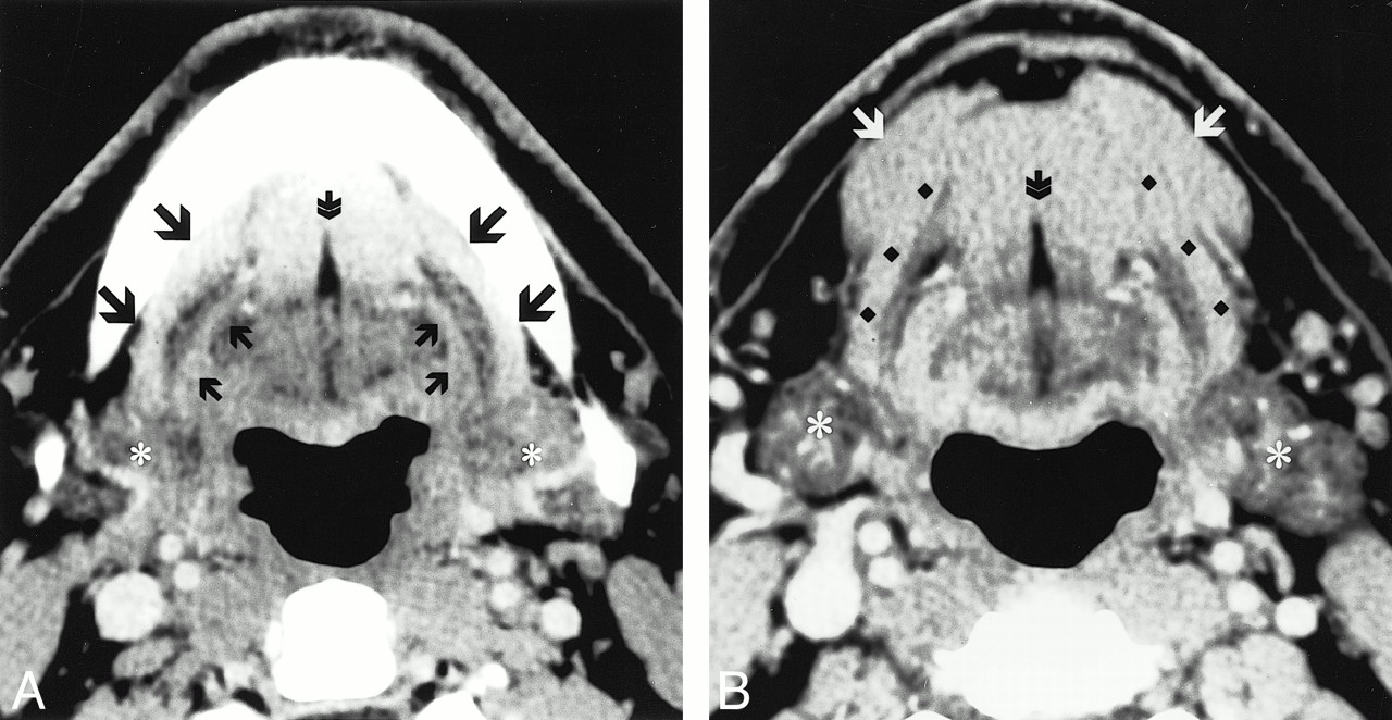
    Normal postcontrast axial CT images through the oral cavity. B is caudal to image A. The classic anatomy of the mylohyoid muscle (A, large black arrows: B, black diamonds) is seen forming a continuous muscular barrier separating the deep sublingual space from the superficial SMS. Running parallel to the posterior aspect of the mylohyoid muscle is the hyoglossus muscle (small black arrows). On either side of the fibro-fatty lingual septum (double arrow) are the genioglossus muscles cranially, and the geniohyoid muscles caudally. Caudally, the anterior belly of the diagastric muscle (white arrows) blends with the superficial margin of the mylohyoid muscle. Asterisks signify the submandibular glands (18)

  • fig 2.
    • Download figure
    • Open in new tab
    • Download powerpoint
    fig 2.

     Postcontrast axial CT images through the oral cavity showing multiple mylohyoid muscle defects. Image A is cranial to image B. On the patients left side, there are two mylohyoid muscle defects. On the right, there is one defect. Accessory salivary tissue is identified bilaterally (large arrows). Note that the accessory salivary tissue has the same attenuation as that of the normal submandibular glands (*).A, large arrow, accessory salivary tissue; small arrows, fat-containing defects. B, large arrows, accessory salivary tissue; small arrow, fat-containing defect (18).

  • fig 3.
    • Download figure
    • Open in new tab
    • Download powerpoint
    fig 3.

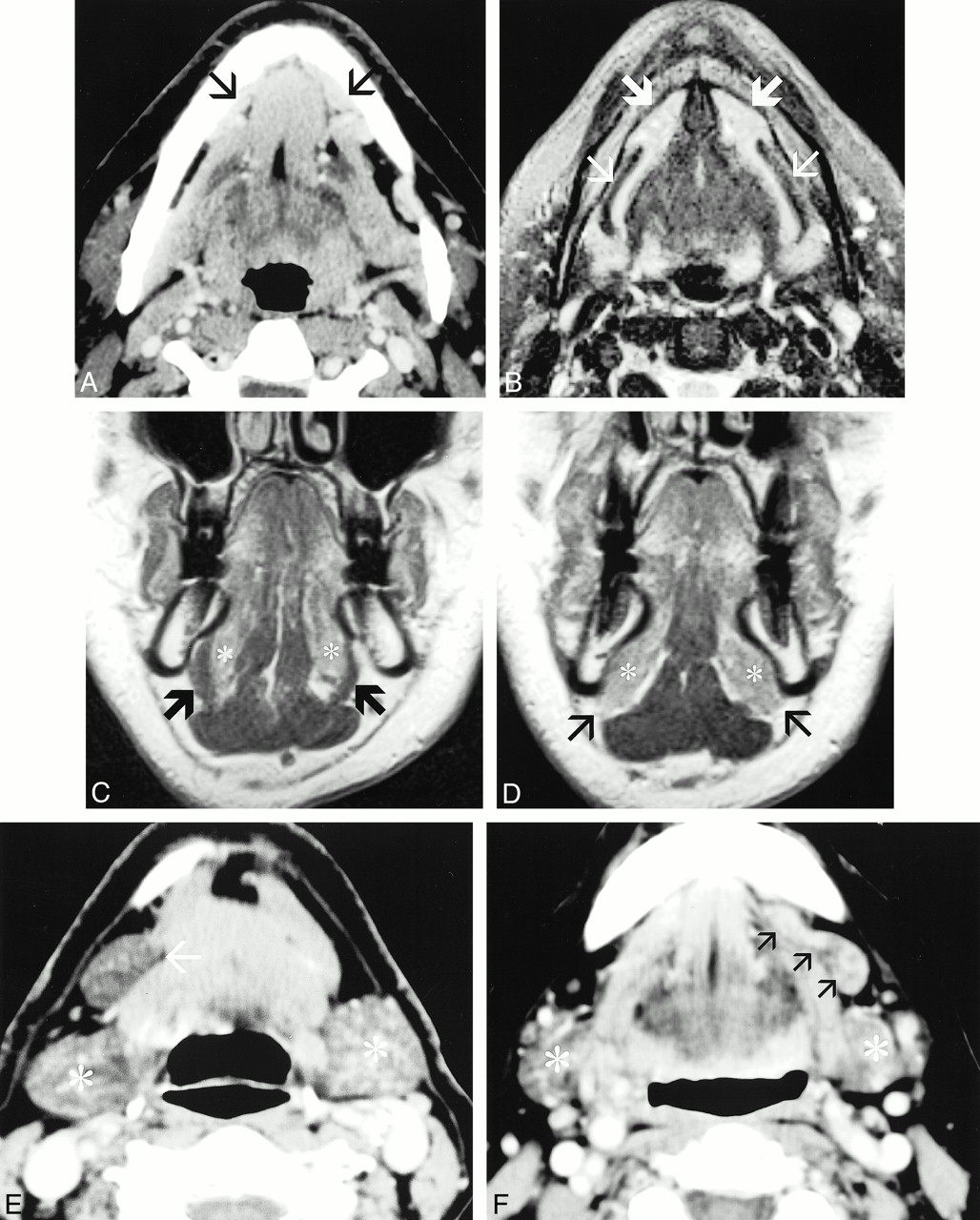
    Accessory salivary tissue has a variable appearance and location in the oral cavity. General features include tissue that is associated with mylohyoid defects and is of similar attenuation and appearance as orthotopic salivary tissue.

    A–D, CT and MR correlation in the same patient with anterior accessory salivary tissue. A, Postcontrast axial CT scan. B, Axial fast spin-echo fat-saturated T2-weighted image. C and D, Postcontrast coronal T1-weighted image. Image C is posterior to image D. The postcontrast CT scan shows enhancing tissue that extends into the submandibular space through deficiencies in the anterior mylohyoid muscle bilaterally (small black arrows). The deficient mylohyoid muscle is clearly identified on the fat-saturated T2-weighted sequence (small white arrows). The normal hyperintense sublingual salivary tissue is clearly identified, extending into the submandibular space anteriorly (large white arrows). The coronal sequences are very useful for identifying the mylohyoid muscle. C depicts the intact mylohyoid muscle (large black arrows) separating the sublingual space and sublingual salivary tissue (*) from the submandibular space. D is located more anteriorly at the level of the mylohyoid defects. Notice the absence of the mylohyoid muscle at this level allows extension of the sublingual salivary tissue into the submandibular space (small black arrows).

    E, Postcontrast CT shows a large deposit of salivary tissue that is located predominately in the submandibular space, with a tapering configuration toward a mid-mylohyoid defect (single arrow). The attenuation and appearance is very similar to the normal submandibular glands (*).

    fig. 3 (Continued)

    F, Postcontrast CT shows accessory salivary tissue, which spans a defect in the mid aspect of the mylohyoid muscle. The tissue is well defined, elongated in configuration, and located in both the sublingual and submandibular space. The attenuation and appearance is very similar to that of the normal submandibular gland (*) (18).

  • fig 4.
    • Download figure
    • Open in new tab
    • Download powerpoint
    fig 4.

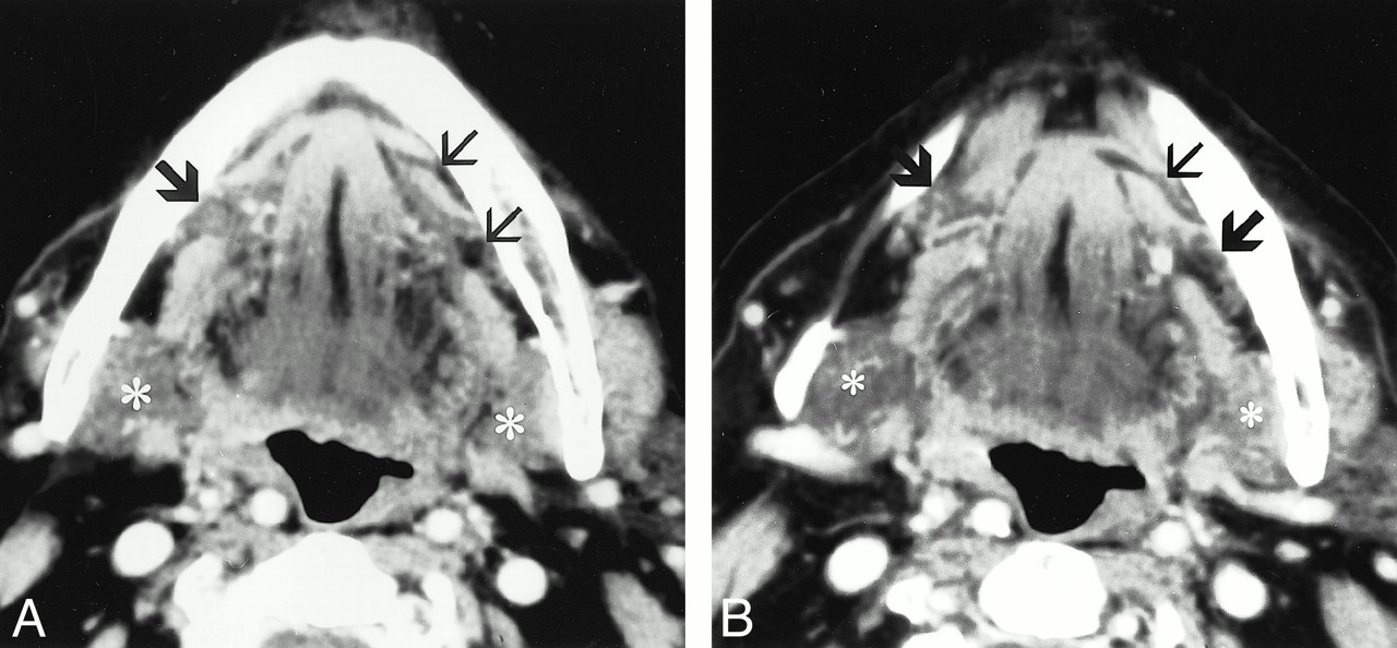
    The most common contents of mylohyoid defects are fat, followed by blood vessels. Fat-containing defects are seen as discrete areas of discontinuity in the mylohyoid muscle, containing low-density adipose tissue. Blood vessels may be identified passing through discrete mylohyoid defects or may be seen perforating the mylohyoid muscle, without an identifiable defect.

    A and B, Postcontrast CT images in different patients. A, On the patient's right side, there is a discrete, wedge-shaped defect in the mid-to-anterior aspect of the mylohyoid muscle (small arrow). On the patient's left side, there is a blood vessel perforating the mylohoid muscle, without an associated defect (large arrow). B, On the patient's left side, there is a large defect in the anterior half of the mylohyoid muscle, containing both fat and a large blood vessel (arrow).

    C and D, CT and coronal CT reformation correlation in the same patient. C is a postcontrast axial CT scan, and D is a coronal, reformatted CT image. These images show a discrete, fat-containing defect in the mid mylohyoid muscle (large arrow). On the coronal image, the small arrow on the patient's left side identifies the intact mylohyoid muscle for comparison (18).

  • fig 5.
    • Download figure
    • Open in new tab
    • Download powerpoint
    fig 5.

    This line drawing illustrates the superficial anatomy of the submandibular space with mylohyoid defects containing accessory salivary tissue and perforating submental branches of the facial artery and vein.

    Large arrow, Accessory salivary tissue; small arrow, mylohyoid defects; large arrowhead, submental branch of facial vein; small arrowhead, submental branch of facial artery; D, anterior belly of diagastric muscle; M, mylohyoid muscle; FA, facial artery off the external carotid artery; FV, facial vein; A, common carotid artery; V, internal jugular vein (18).

Tables

  • Figures
  • Table1

    One hundred individuals scanned

PreviousNext
Back to top

In this issue

American Journal of Neuroradiology
Vol. 22, Issue 2
1 Feb 2001
  • Table of Contents
  • Index by author
Advertisement
Print
Download PDF
Email Article

Thank you for your interest in spreading the word on American Journal of Neuroradiology.

NOTE: We only request your email address so that the person you are recommending the page to knows that you wanted them to see it, and that it is not junk mail. We do not capture any email address.

Enter multiple addresses on separate lines or separate them with commas.
Accessory Salivary Tissue in the Mylohyoid Boutonnière: A Clinical and Radiologic Pseudolesion of the Oral Cavity
(Your Name) has sent you a message from American Journal of Neuroradiology
(Your Name) thought you would like to see the American Journal of Neuroradiology web site.
CAPTCHA
This question is for testing whether or not you are a human visitor and to prevent automated spam submissions.
Cite this article
David K. White, H. Christian Davidson, H. Ric Harnsberger, Jeffrey Haller, Aya Kamya
Accessory Salivary Tissue in the Mylohyoid Boutonnière: A Clinical and Radiologic Pseudolesion of the Oral Cavity
American Journal of Neuroradiology Feb 2001, 22 (2) 406-412;

Citation Manager Formats

  • BibTeX
  • Bookends
  • EasyBib
  • EndNote (tagged)
  • EndNote 8 (xml)
  • Medlars
  • Mendeley
  • Papers
  • RefWorks Tagged
  • Ref Manager
  • RIS
  • Zotero
0 Responses
Respond to this article
Share
Bookmark this article
Accessory Salivary Tissue in the Mylohyoid Boutonnière: A Clinical and Radiologic Pseudolesion of the Oral Cavity
David K. White, H. Christian Davidson, H. Ric Harnsberger, Jeffrey Haller, Aya Kamya
American Journal of Neuroradiology Feb 2001, 22 (2) 406-412;
del.icio.us logo Twitter logo Facebook logo Mendeley logo
  • Tweet Widget
  • Facebook Like
  • Google Plus One

Jump to section

  • Article
    • Abstract
    • Methods
    • Results
    • Discussion
    • Conclusion
    • Footnotes
    • References
  • Figures & Data
  • Info & Metrics
  • Responses
  • References
  • PDF

Related Articles

  • No related articles found.
  • PubMed
  • Google Scholar

Cited By...

  • Types of Sublingual Gland Herniation Observed During Sonography of Plunging Ranulas
  • CT and MR findings of bilateral submandibular gland aplasia associated with hypertrophied symmetrical sublingual glands herniated through mylohyoid defects
  • Imaging the oral cavity: key concepts for the radiologist
  • Bilateral submandibular gland aplasia with clinico-radiological mass due to prolapsing sublingual salivary tissue through mylohyoid boutonniere: a case report and review
  • Mylohyoid herniation of the sublingual gland diagnosed by magnetic resonance imaging
  • Giant Ranula of the Neck: Differentiation from Cystic Hygroma
  • Crossref
  • Google Scholar

This article has not yet been cited by articles in journals that are participating in Crossref Cited-by Linking.

More in this TOC Section

  • FDG Uptake and Thyroid Cancer Post-BRAF Therapy
  • Normal Facial Nerve Enhancement on VIBE Sequence
  • Contrast Enhanced Pituitary CISS/FIESTA
Show more HEAD AND NECK IMAGING

Similar Articles

Advertisement

Indexed Content

  • Current Issue
  • Accepted Manuscripts
  • Article Preview
  • Past Issues
  • Editorials
  • Editor's Choice
  • Fellows' Journal Club
  • Letters to the Editor
  • Video Articles

Cases

  • Case Collection
  • Archive - Case of the Week
  • Archive - Case of the Month
  • Archive - Classic Case

More from AJNR

  • Trainee Corner
  • Imaging Protocols
  • MRI Safety Corner
  • Book Reviews

Multimedia

  • AJNR Podcasts
  • AJNR Scantastics

Resources

  • Turnaround Time
  • Submit a Manuscript
  • Submit a Video Article
  • Submit an eLetter to the Editor/Response
  • Manuscript Submission Guidelines
  • Statistical Tips
  • Fast Publishing of Accepted Manuscripts
  • Graphical Abstract Preparation
  • Imaging Protocol Submission
  • Evidence-Based Medicine Level Guide
  • Publishing Checklists
  • Author Policies
  • Become a Reviewer/Academy of Reviewers
  • News and Updates

About Us

  • About AJNR
  • Editorial Board
  • Editorial Board Alumni
  • Alerts
  • Permissions
  • Not an AJNR Subscriber? Join Now
  • Advertise with Us
  • Librarian Resources
  • Feedback
  • Terms and Conditions
  • AJNR Editorial Board Alumni

American Society of Neuroradiology

  • Not an ASNR Member? Join Now

© 2025 by the American Society of Neuroradiology All rights, including for text and data mining, AI training, and similar technologies, are reserved.
Print ISSN: 0195-6108 Online ISSN: 1936-959X

Powered by HighWire