
Article Figures & Data
Figures
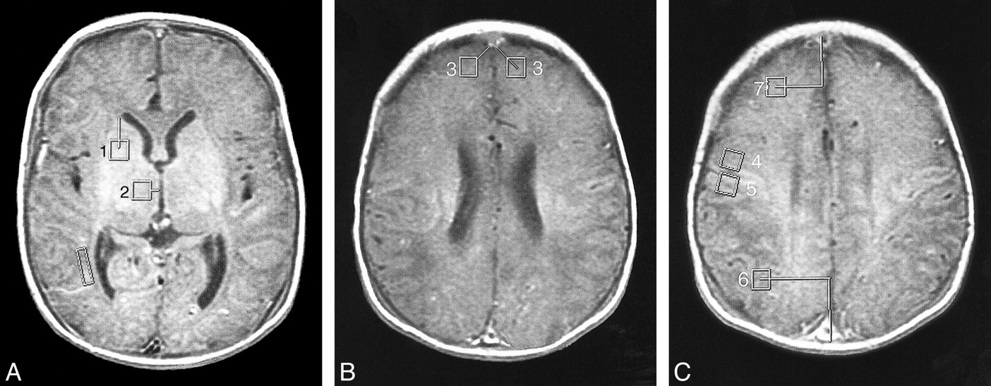
- fig 1.
Locations of the 40-mm2 voxels used in the study (see Table 1). The location of the eyeball voxel is obvious and not illustrated. The lines show how the measurements were made to keep the locations consistent.
A, MR image shows location of voxels for basal ganglia (1) and thalamus (2). (The box adjacent to the trigone/occipital horn of the right lateral ventricle was for another project.)
B, MR image shows location of voxels for frontal white matter (3).
C, MR image shows location of voxels for prerolandic (4), postrolandic (5), posterior watershed (6), and anterior watershed (7) areas.

Tables

- TABLE 2:
Quantitative analysis A: Results from stepwise logistic regression using quantitative MR variables
- TABLE 2:
B: Results from stepwise linear regression using quantitative MR variables
- TABLE 2:
C: Results from stepwise linear regression using quantitative MR variables
- TABLE 3:
Qualitative Analysis A: Results from stepwise logistic regression using qualitative MR variables
- TABLE 3:
B: Results from stepwise linear regression using qualitative MR variables
- TABLE 3:
C: Results from stepwise linear regression using qualitative MR variables


