
Article Figures & Data
Figures
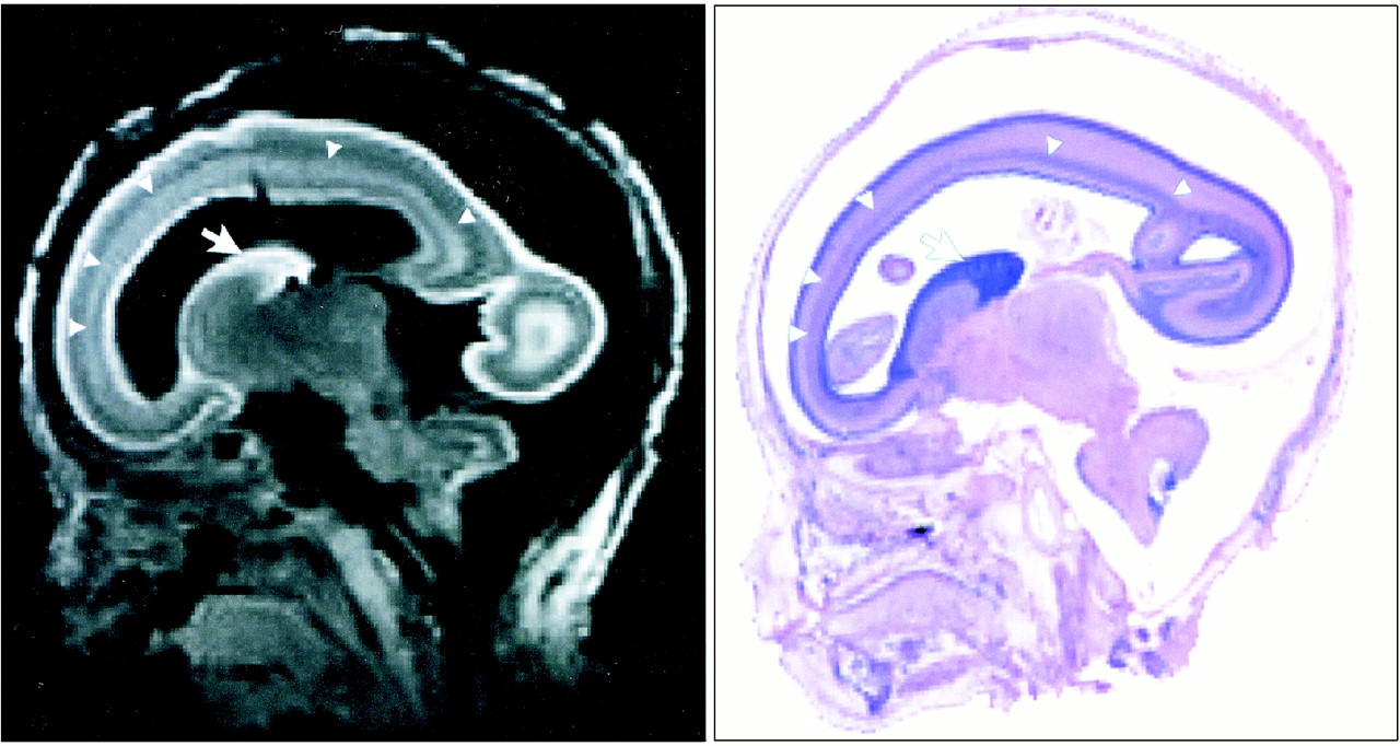
- fig 1.
T1-weighted MR image (left) reconstructed from 3D-SSFP sequence and sagittal H&E-stained photomicrograph (right) of fetal brain at 21 weeks' GA. There is good correlation between the images. Each image reveals the germinal matrix (arrows) and migrating neuroblast layer (arrowheads)
- fig 2.
Images show good correlation between macroscopic brain (upper row) and 3D surface-rendered brain (middle row). Germinal matrix (orange), located ventrolateral to the lateral ventricles (blue), extends along the lateral walls of the lateral ventricles (lower row).
- fig 3.
Successive images show developmental changes of lateral configuration of brain, germinal matrix, and ventricular system. The brain surfaces (upper row), germinal matrix (middle row, orange), and ventricular system (lower row, blue) of human fetal brain were reconstructed by surface rendering. The volume of the germinal matrix increased until 23 weeks' GA and decreased rapidly at 28 weeks' GA. Note how lateral ventricles change from fetal type, with vesicular aspect and bicornuate shape, to adult type with increasing GA.
- fig 4.
Serial coronal MR images of the fetus at 7 weeks' GA. Germinal matrix cannot be detected
- fig 5.
Serial coronal MR images of the fetus at 9 weeks' GA. Note germinal matrix is detected ventrolateral to the lateral ventricles (arrow)
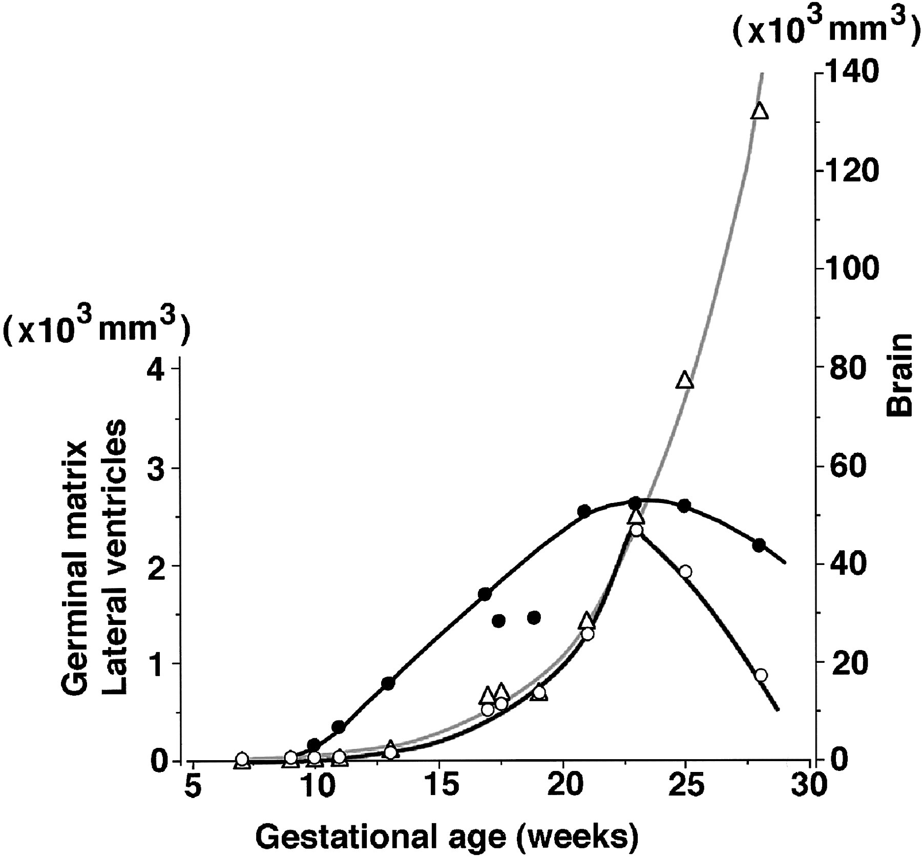
- fig 6.
Volumetric changes of the fetal brain (open triangles), germinal matrix (open circles), and lateral ventricles (solid circles). Increasing fetal brain volume (gray curve) has an exponential relationship (r2 = .963) to GA, reaching 132.5 cm3 at 28 weeks' GA. The germinal matrix also increases exponentially, reaching a volume of 2.3 cm3 at 23 weeks' GA, then decreases rapidly after 25 weeks' GA. In contrast to the germinal matrix, the volume of the lateral ventricles gradually increases, up to 2.6 cm3 at 23 weeks' GA
- fig 7.
Relationship between germinal matrix (open circles), lateral ventricles (solid circles), and brain. It is noteworthy that the volumetric ratio of the germinal matrix to brain volume is constant at about 5% between 11 and 23 weeks' GA


In this issue


Jump to section
Related Articles
Cited By...
- Fetal Brain Growth in the Early Second Trimester
- Three-dimensional cranial ultrasound and functional near infrared spectroscopy for bedside monitoring of intraventricular hemorrhage in preterm neonates
- The Forget-Me-Not dHCP study: 7 Tesla high resolution diffusion imaging in the unfixed post-mortem neonatal brain
- The Japan Monkey Centre Primates Brain Imaging Repository of high-resolution postmortem magnetic resonance imaging: the second phase of the archive of digital records
- The Lateral Ventricles: A Detailed Review of Anatomy, Development, and Anatomic Variations
- Morphologic Evolution and Coordinated Development of the Fetal Lateral Ventricles in the Second and Third Trimesters
- Characterization of the ventricular-subventricular stem cell niche during human brain development
- MR Imaging of the Pituitary Gland and Postsphenoid Ossification in Fetal Specimens
- Magnetic Resonance Volumetric Assessments of Brains in Fetuses With Ventriculomegaly Correlated to Outcomes
- Local Tissue Growth Patterns Underlying Normal Fetal Human Brain Gyrification Quantified In Utero
- Prospective parental consent for autopsy research following sudden unexpected childhood deaths: a successful model
- Anatomical Characterization of Human Fetal Brain Development with Diffusion Tensor Magnetic Resonance Imaging