Advertisement

Main menu

  • Home
  • Content
    • Current Issue
    • Accepted Manuscripts
    • Article Preview
    • Past Issue Archive
    • Video Articles
    • AJNR Case Collection
    • Case of the Week Archive
    • Case of the Month Archive
    • Classic Case Archive
  • Special Collections
    • AJNR Awards
    • Low-Field MRI
    • Alzheimer Disease
    • ASNR Foundation Special Collection
    • Photon-Counting CT
    • View All
  • Multimedia
    • AJNR Podcasts
    • AJNR SCANtastic
    • Trainee Corner
    • MRI Safety Corner
    • Imaging Protocols
  • For Authors
    • Submit a Manuscript
    • Submit a Video Article
    • Submit an eLetter to the Editor/Response
    • Manuscript Submission Guidelines
    • Statistical Tips
    • Fast Publishing of Accepted Manuscripts
    • Graphical Abstract Preparation
    • Imaging Protocol Submission
    • Author Policies
  • About Us
    • About AJNR
    • Editorial Board
    • Editorial Board Alumni
  • More
    • Become a Reviewer/Academy of Reviewers
    • Subscribers
    • Permissions
    • Alerts
    • Feedback
    • Advertisers
    • ASNR Home

User menu

  • Alerts
  • Log in

Search

  • Advanced search
American Journal of Neuroradiology

American Journal of Neuroradiology

ASHNR American Society of Functional Neuroradiology ASHNR American Society of Pediatric Neuroradiology ASSR
  • Alerts
  • Log in

Advanced Search

  • Home
  • Content
    • Current Issue
    • Accepted Manuscripts
    • Article Preview
    • Past Issue Archive
    • Video Articles
    • AJNR Case Collection
    • Case of the Week Archive
    • Case of the Month Archive
    • Classic Case Archive
  • Special Collections
    • AJNR Awards
    • Low-Field MRI
    • Alzheimer Disease
    • ASNR Foundation Special Collection
    • Photon-Counting CT
    • View All
  • Multimedia
    • AJNR Podcasts
    • AJNR SCANtastic
    • Trainee Corner
    • MRI Safety Corner
    • Imaging Protocols
  • For Authors
    • Submit a Manuscript
    • Submit a Video Article
    • Submit an eLetter to the Editor/Response
    • Manuscript Submission Guidelines
    • Statistical Tips
    • Fast Publishing of Accepted Manuscripts
    • Graphical Abstract Preparation
    • Imaging Protocol Submission
    • Author Policies
  • About Us
    • About AJNR
    • Editorial Board
    • Editorial Board Alumni
  • More
    • Become a Reviewer/Academy of Reviewers
    • Subscribers
    • Permissions
    • Alerts
    • Feedback
    • Advertisers
    • ASNR Home
  • Follow AJNR on Twitter
  • Visit AJNR on Facebook
  • Follow AJNR on Instagram
  • Join AJNR on LinkedIn
  • RSS Feeds

AJNR Awards, New Junior Editors, and more. Read the latest AJNR updates

Research ArticleBRAIN

An Adult Case of Congenital External Carotid–Jugular Arteriovenous Fistula with Reversible Circulatory Insufficiency in the Cerebellum and Lower Brain Stem

Masahiro Horiuchi, Tsutomu Kamo, Hiroshi Sugihara, Koshiro Fujisawa, Yoichi Takahashi, Ritsuko Ikeda, Hiroyuki Morishima and Yoshitaro Yamaguchi
American Journal of Neuroradiology February 2001, 22 (2) 273-276;
Masahiro Horiuchi
  • Find this author on Google Scholar
  • Find this author on PubMed
  • Search for this author on this site
Tsutomu Kamo
  • Find this author on Google Scholar
  • Find this author on PubMed
  • Search for this author on this site
Hiroshi Sugihara
  • Find this author on Google Scholar
  • Find this author on PubMed
  • Search for this author on this site
Koshiro Fujisawa
  • Find this author on Google Scholar
  • Find this author on PubMed
  • Search for this author on this site
Yoichi Takahashi
  • Find this author on Google Scholar
  • Find this author on PubMed
  • Search for this author on this site
Ritsuko Ikeda
  • Find this author on Google Scholar
  • Find this author on PubMed
  • Search for this author on this site
Hiroyuki Morishima
  • Find this author on Google Scholar
  • Find this author on PubMed
  • Search for this author on this site
Yoshitaro Yamaguchi
  • Find this author on Google Scholar
  • Find this author on PubMed
  • Search for this author on this site
  • Article
  • Figures & Data
  • Info & Metrics
  • Responses
  • References
  • PDF
Loading

Article Figures & Data

Figures

  • fig 1.
    • Download figure
    • Open in new tab
    • Download powerpoint
    fig 1.

    27-year-old man with congenital external carotid–jugular arteriovenous fistula who presented with a diminished level of consciousness and an ataxic gait.

    A, Axial FLAIR MR images (6000/120) on the day of hospital admission show venous congestion, a dilated right jugular vein, and an area of high signal intensity in the brain stem and cerebellum. A small infarction is visible in the temporal lobe.

    B and C, Angiograms, frontal (B) and lateral (C) views, 1 day after admission show a dilated right external carotid artery and internal jugular vein and the presence of a fistula.

    D, Brain CT studies 5 hours after angiography show congestion and edema.
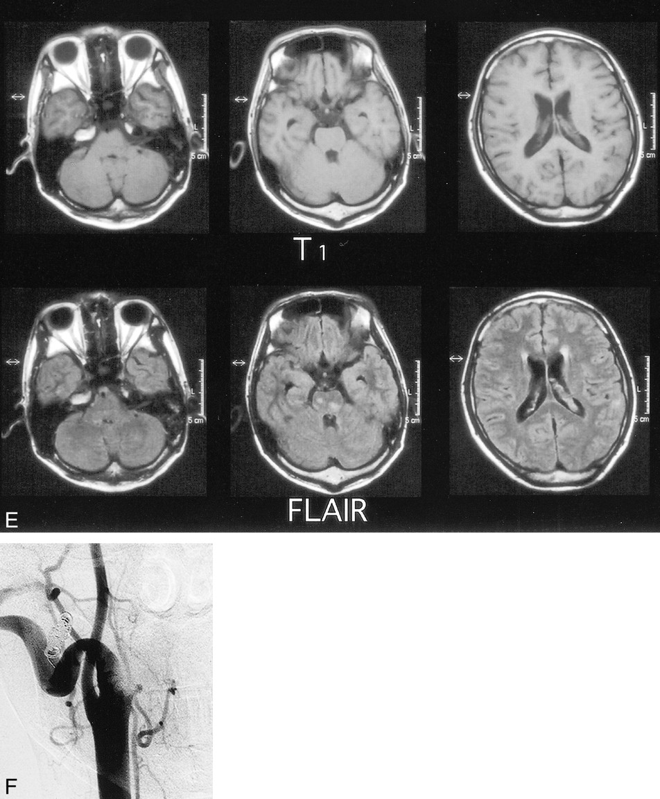
  • fig 1.
    • Download figure
    • Open in new tab
    • Download powerpoint
    fig 1.

    fig. 1 Continued.

    E, Axial FLAIR MR images (6000/120) after embolization show only a few areas of high signal intensity in the brain stem and cerebellum.

    F, Angiogram 1 month after admission shows that the external carotid artery and jugular vein are almost equivalent in size, and no arteriovenous communication is detected.

PreviousNext
Back to top

In this issue

American Journal of Neuroradiology
Vol. 22, Issue 2
1 Feb 2001
  • Table of Contents
  • Index by author
Advertisement
Print
Download PDF
Email Article

Thank you for your interest in spreading the word on American Journal of Neuroradiology.

NOTE: We only request your email address so that the person you are recommending the page to knows that you wanted them to see it, and that it is not junk mail. We do not capture any email address.

Enter multiple addresses on separate lines or separate them with commas.
An Adult Case of Congenital External Carotid–Jugular Arteriovenous Fistula with Reversible Circulatory Insufficiency in the Cerebellum and Lower Brain Stem
(Your Name) has sent you a message from American Journal of Neuroradiology
(Your Name) thought you would like to see the American Journal of Neuroradiology web site.
CAPTCHA
This question is for testing whether or not you are a human visitor and to prevent automated spam submissions.
Cite this article
Masahiro Horiuchi, Tsutomu Kamo, Hiroshi Sugihara, Koshiro Fujisawa, Yoichi Takahashi, Ritsuko Ikeda, Hiroyuki Morishima, Yoshitaro Yamaguchi
An Adult Case of Congenital External Carotid–Jugular Arteriovenous Fistula with Reversible Circulatory Insufficiency in the Cerebellum and Lower Brain Stem
American Journal of Neuroradiology Feb 2001, 22 (2) 273-276;

Citation Manager Formats

  • BibTeX
  • Bookends
  • EasyBib
  • EndNote (tagged)
  • EndNote 8 (xml)
  • Medlars
  • Mendeley
  • Papers
  • RefWorks Tagged
  • Ref Manager
  • RIS
  • Zotero
0 Responses
Respond to this article
Share
Bookmark this article
An Adult Case of Congenital External Carotid–Jugular Arteriovenous Fistula with Reversible Circulatory Insufficiency in the Cerebellum and Lower Brain Stem
Masahiro Horiuchi, Tsutomu Kamo, Hiroshi Sugihara, Koshiro Fujisawa, Yoichi Takahashi, Ritsuko Ikeda, Hiroyuki Morishima, Yoshitaro Yamaguchi
American Journal of Neuroradiology Feb 2001, 22 (2) 273-276;
del.icio.us logo Twitter logo Facebook logo Mendeley logo
  • Tweet Widget
  • Facebook Like
  • Google Plus One

Jump to section

  • Article
    • Abstract
    • Case Report
    • Discussion
    • Footnotes
    • References
  • Figures & Data
  • Info & Metrics
  • Responses
  • References
  • PDF

Related Articles

  • No related articles found.
  • PubMed
  • Google Scholar

Cited By...

  • No citing articles found.
  • Crossref
  • Google Scholar

This article has not yet been cited by articles in journals that are participating in Crossref Cited-by Linking.

More in this TOC Section

  • Multimodal CT Provides Improved Performance for Lacunar Infarct Detection
  • Optimal MRI Sequence for Identifying Occlusion Location in Acute Stroke: Which Value of Time-Resolved Contrast-Enhanced MRA?
  • Evaluating the Effects of White Matter Multiple Sclerosis Lesions on the Volume Estimation of 6 Brain Tissue Segmentation Methods
Show more BRAIN

Similar Articles

Advertisement

Indexed Content

  • Current Issue
  • Accepted Manuscripts
  • Article Preview
  • Past Issues
  • Editorials
  • Editor's Choice
  • Fellows' Journal Club
  • Letters to the Editor
  • Video Articles

Cases

  • Case Collection
  • Archive - Case of the Week
  • Archive - Case of the Month
  • Archive - Classic Case

More from AJNR

  • Trainee Corner
  • Imaging Protocols
  • MRI Safety Corner

Multimedia

  • AJNR Podcasts
  • AJNR Scantastics

Resources

  • Turnaround Time
  • Submit a Manuscript
  • Submit a Video Article
  • Submit an eLetter to the Editor/Response
  • Manuscript Submission Guidelines
  • Statistical Tips
  • Fast Publishing of Accepted Manuscripts
  • Graphical Abstract Preparation
  • Imaging Protocol Submission
  • Evidence-Based Medicine Level Guide
  • Publishing Checklists
  • Author Policies
  • Become a Reviewer/Academy of Reviewers
  • News and Updates

About Us

  • About AJNR
  • Editorial Board
  • Editorial Board Alumni
  • Alerts
  • Permissions
  • Not an AJNR Subscriber? Join Now
  • Advertise with Us
  • Librarian Resources
  • Feedback
  • Terms and Conditions
  • AJNR Editorial Board Alumni

American Society of Neuroradiology

  • Not an ASNR Member? Join Now

© 2025 by the American Society of Neuroradiology All rights, including for text and data mining, AI training, and similar technologies, are reserved.
Print ISSN: 0195-6108 Online ISSN: 1936-959X

Powered by HighWire