Advertisement
  • Main menu
  • User menu
  • Search
  • English ▼
    • English
    • Afrikaans
    • Albanian
    • Amharic
    • Arabic
    • Armenian
    • Azerbaijani
    • Basque
    • Belarusian
    • Bengali
    • Bosnian
    • Bulgarian
    • Catalan
    • Cebuano
    • Chichewa
    • Chinese (Simplified)
    • Chinese (Traditional)
    • Corsican
    • Croatian
    • Czech
    • Danish
    • Dutch
    • Esperanto
    • Estonian
    • Filipino
    • Finnish
    • French
    • Frisian
    • Galician
    • Georgian
    • German
    • Greek
    • Gujarati
    • Haitian Creole
    • Hausa
    • Hawaiian
    • Hebrew
    • Hindi
    • Hmong
    • Hungarian
    • Icelandic
    • Igbo
    • Indonesian
    • Irish
    • Italian
    • Japanese
    • Javanese
    • Kannada
    • Kazakh
    • Khmer
    • Korean
    • Kurdish (Kurmanji)
    • Kyrgyz
    • Lao
    • Latin
    • Latvian
    • Lithuanian
    • Luxembourgish
    • Macedonian
    • Malagasy
    • Malay
    • Malayalam
    • Maltese
    • Maori
    • Marathi
    • Mongolian
    • Myanmar (Burmese)
    • Nepali
    • Norwegian
    • Pashto
    • Persian
    • Polish
    • Portuguese
    • Punjabi
    • Romanian
    • Russian
    • Samoan
    • Scottish Gaelic
    • Serbian
    • Sesotho
    • Shona
    • Sindhi
    • Sinhala
    • Slovak
    • Slovenian
    • Somali
    • Spanish
    • Sudanese
    • Swahili
    • Swedish
    • Tajik
    • Tamil
    • Telugu
    • Thai
    • Turkish
    • Ukrainian
    • Urdu
    • Uzbek
    • Vietnamese
    • Welsh
    • Xhosa
    • Yiddish
    • Yoruba
    • Zulu

Main menu

  • Home
  • Content
    • Current Issue
    • Accepted Manuscripts
    • Article Preview
    • Past Issue Archive
    • Video Articles
    • AJNR Case Collection
    • Case of the Week Archive
    • Case of the Month Archive
    • Classic Case Archive
  • Special Collections
    • Advancing NeuroMRI with High-Relaxivity Contrast Agents
    • AJNR Awards
    • Low-Field MRI
    • Alzheimer Disease
    • ASNR Foundation Special Collection
    • Photon-Counting CT
    • View All
  • Multimedia
    • AJNR Podcasts
    • AJNR SCANtastic
    • Trainee Corner
    • MRI Safety Corner
    • Imaging Protocols
  • For Authors
    • Submit a Manuscript
    • Submit a Video Article
    • Submit an eLetter to the Editor/Response
    • Manuscript Submission Guidelines
    • Statistical Tips
    • Fast Publishing of Accepted Manuscripts
    • Graphical Abstract Preparation
    • Imaging Protocol Submission
    • Author Policies
  • About Us
    • About AJNR
    • Editorial Board
    • Editorial Board Alumni
  • More
    • Become a Reviewer/Academy of Reviewers
    • Subscribers
    • Permissions
    • Alerts
    • Feedback
    • Advertisers
    • ASNR Home

User menu

  • Alerts
  • Log in

Search

  • Advanced search
American Journal of Neuroradiology

American Journal of Neuroradiology

ASHNR American Society of Functional Neuroradiology ASHNR American Society of Pediatric Neuroradiology ASSR
  • Alerts
  • Log in

Advanced Search

English ▼
  • English
  • Afrikaans
  • Albanian
  • Amharic
  • Arabic
  • Armenian
  • Azerbaijani
  • Basque
  • Belarusian
  • Bengali
  • Bosnian
  • Bulgarian
  • Catalan
  • Cebuano
  • Chichewa
  • Chinese (Simplified)
  • Chinese (Traditional)
  • Corsican
  • Croatian
  • Czech
  • Danish
  • Dutch
  • Esperanto
  • Estonian
  • Filipino
  • Finnish
  • French
  • Frisian
  • Galician
  • Georgian
  • German
  • Greek
  • Gujarati
  • Haitian Creole
  • Hausa
  • Hawaiian
  • Hebrew
  • Hindi
  • Hmong
  • Hungarian
  • Icelandic
  • Igbo
  • Indonesian
  • Irish
  • Italian
  • Japanese
  • Javanese
  • Kannada
  • Kazakh
  • Khmer
  • Korean
  • Kurdish (Kurmanji)
  • Kyrgyz
  • Lao
  • Latin
  • Latvian
  • Lithuanian
  • Luxembourgish
  • Macedonian
  • Malagasy
  • Malay
  • Malayalam
  • Maltese
  • Maori
  • Marathi
  • Mongolian
  • Myanmar (Burmese)
  • Nepali
  • Norwegian
  • Pashto
  • Persian
  • Polish
  • Portuguese
  • Punjabi
  • Romanian
  • Russian
  • Samoan
  • Scottish Gaelic
  • Serbian
  • Sesotho
  • Shona
  • Sindhi
  • Sinhala
  • Slovak
  • Slovenian
  • Somali
  • Spanish
  • Sudanese
  • Swahili
  • Swedish
  • Tajik
  • Tamil
  • Telugu
  • Thai
  • Turkish
  • Ukrainian
  • Urdu
  • Uzbek
  • Vietnamese
  • Welsh
  • Xhosa
  • Yiddish
  • Yoruba
  • Zulu
  • Home
  • Content
    • Current Issue
    • Accepted Manuscripts
    • Article Preview
    • Past Issue Archive
    • Video Articles
    • AJNR Case Collection
    • Case of the Week Archive
    • Case of the Month Archive
    • Classic Case Archive
  • Special Collections
    • Advancing NeuroMRI with High-Relaxivity Contrast Agents
    • AJNR Awards
    • Low-Field MRI
    • Alzheimer Disease
    • ASNR Foundation Special Collection
    • Photon-Counting CT
    • View All
  • Multimedia
    • AJNR Podcasts
    • AJNR SCANtastic
    • Trainee Corner
    • MRI Safety Corner
    • Imaging Protocols
  • For Authors
    • Submit a Manuscript
    • Submit a Video Article
    • Submit an eLetter to the Editor/Response
    • Manuscript Submission Guidelines
    • Statistical Tips
    • Fast Publishing of Accepted Manuscripts
    • Graphical Abstract Preparation
    • Imaging Protocol Submission
    • Author Policies
  • About Us
    • About AJNR
    • Editorial Board
    • Editorial Board Alumni
  • More
    • Become a Reviewer/Academy of Reviewers
    • Subscribers
    • Permissions
    • Alerts
    • Feedback
    • Advertisers
    • ASNR Home
  • Follow AJNR on Twitter
  • Visit AJNR on Facebook
  • Follow AJNR on Instagram
  • Join AJNR on LinkedIn
  • RSS Feeds

AJNR Awards, New Junior Editors, and more. Read the latest AJNR updates


Improved Turnaround Times | Median time to first decision: 12 days

Research ArticlePediatric Neuroimaging

MR Findings in Spinal Hemangioblastoma: Correlation with Symptoms and with Angiographic and Surgical Findings

Bao-Cheng Chu, Satoshi Terae, Kazutoshi Hida, Matakazu Furukawa, Satoru Abe and Kazuo Miyasaka
American Journal of Neuroradiology January 2001, 22 (1) 206-217;
Bao-Cheng Chu
  • Find this author on Google Scholar
  • Find this author on PubMed
  • Search for this author on this site
Satoshi Terae
  • Find this author on Google Scholar
  • Find this author on PubMed
  • Search for this author on this site
Kazutoshi Hida
  • Find this author on Google Scholar
  • Find this author on PubMed
  • Search for this author on this site
Matakazu Furukawa
  • Find this author on Google Scholar
  • Find this author on PubMed
  • Search for this author on this site
Satoru Abe
  • Find this author on Google Scholar
  • Find this author on PubMed
  • Search for this author on this site
Kazuo Miyasaka
  • Find this author on Google Scholar
  • Find this author on PubMed
  • Search for this author on this site
  • Article
  • Figures & Data
  • Info & Metrics
  • Responses
  • References
  • PDF
Loading

Article Figures & Data

Figures

  • Tables
  • fig 1.
    • Download figure
    • Open in new tab
    • Download powerpoint
    fig 1.

    Case 6: 30-year-old man without VHLD.

    A and B, Sagittal T1-weighted SE images (400/23/2) before (A) and after (B) intravenous administration of contrast material show a large, sausagelike, well-demarcated, intensely but heterogeneously enhancing tumor at T12–L1. Note superficial enhancement of the spinal cord (arrows, B), confirmed to be dilated perimedullary veins at surgery. Cephalic portion of the tumor resides within the spinal cord, and the caudal portion is extramedullary. A large intramedullary tumor with exophytic growth was confirmed at surgery.

    C, Sagittal T2-weighted FSE image (4000/96/3) shows mixed hyper- and isointense tumor. Cephalic to this tumor is syringomyelia up to C2 level (only partially shown). Note prominent vascular flow voids (arrows).

    D and E, Arterial (D) and venous (E) phases of digital subtraction angiograms, anteroposterior view, with right T10 intercostal artery catheterized, show intense tumor stain at T12–L1, dilated posterior spinal artery as a feeder (straight arrows, D), and dilated draining vein (curved arrows, E).
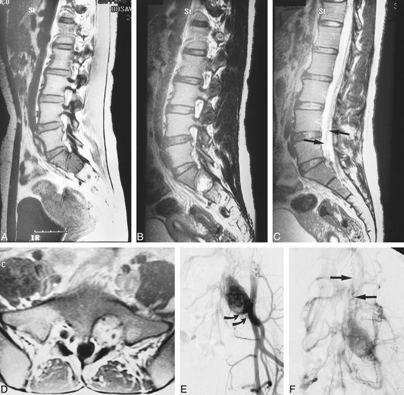
  • fig 2.
    • Download figure
    • Open in new tab
    • Download powerpoint
    fig 2.

    Case 3: 24-year-old woman with VHLD.

    A, Sagittal T1-weighted FSE image (700/10/6) shows a large, round, hypointense tumor in the left vertebral foramen between S1 and S2. Note vascular flow voids within the tumor.

    B, Sagittal T2-weighted FSE image (4100/121/5) shows mixed iso- and hyperintense tumor.

    C, Sagittal T2-weighted FSE image (4100/121/5), medial to slice in B, shows serpentine vascular flow voids (arrows).

    D, Contrast-enhanced axial T1-weighted FSE image (740/8.6/4) shows intensely but heterogeneously enhancing tumor that was confirmed to arise from left S1 nerve root at surgery.

    E and F, Arterial (E) and venous (F) phases of digital subtraction angiograms, right anterior oblique 30° view, with left internal iliac artery catheterized, show intense tumor stain, dilated lateral sacral arteries that supply the tumor (curved arrows, E), and early venous filling (straight arrows, F), indicating the arteriovenous shunt in the tumor. This vein corresponds with the flow voids seen in C.

  • fig 3.
    • Download figure
    • Open in new tab
    • Download powerpoint
    fig 3.

    Case 2: 42-year-old woman with VHLD without subjective symptoms.

    A and B, Contrast-enhanced sagittal T1-weighted FSE images (600/12/6) show nodular, intensely and homogeneously enhancing tumors at the posterior aspects of the spinal cord at levels C5, C6, T8–T9, and T9–T10.

    C, Sagittal T2-weighted TSE image (4500/112/3) shows a syrinx at T7–T8 and adjacent edema at T9–T10. This image is a composite of the upper and lower halves of the MR images of the spine.

    D, Contrast-enhanced axial T1-weighted TSE image (1000/12/2) shows the tumor surrounding a posterior nerve root at C6, as is also the case with other tumors at C5 and C7–T1 (not shown).

  • fig 4.
    • Download figure
    • Open in new tab
    • Download powerpoint
    fig 4.

    Case 3: 24-year-old woman with VHLD.

    A, Sagittal T1-weighted FSE image (700/10/6) shows cord enlargement and decreased signal intensity at T3–T6.

    B, Contrast-enhanced sagittal T1-weighted FSE image (700/10/6) shows a small, ovoid, well-demarcated, intensely and homogeneously enhancing tumor at T4–T5.

    C, Sagittal T2-weighted FSE image (4220/120/5) shows isointense tumor (arrow) and hyperintense pencil-shaped lesion from T3 to T6.

    D, Contrast-enhanced axial T1-weighted FSE image (540/9/3) shows the tumor is superficially located at the posterior aspect of the spinal cord.

  • fig 5.
    • Download figure
    • Open in new tab
    • Download powerpoint
    fig 5.

    Case 4: 44-year-old woman with VHLD who had no spinal cord symptoms.

    A, Contrast-enhanced sagittal T1-weighted SE image (800/14/2) shows a small, ovoid, well-demarcated, intensely enhancing intramedullary tumor at the anterior aspect of the spinal cord at T10.

    B, Sagittal T2-weighted TSE image (4500/112/3) shows edema (arrow).

    C, Contrast-enhanced axial T1-weighted SE image (660/15/1) shows the tumor is superficially located at the anterior aspect of the spinal cord.
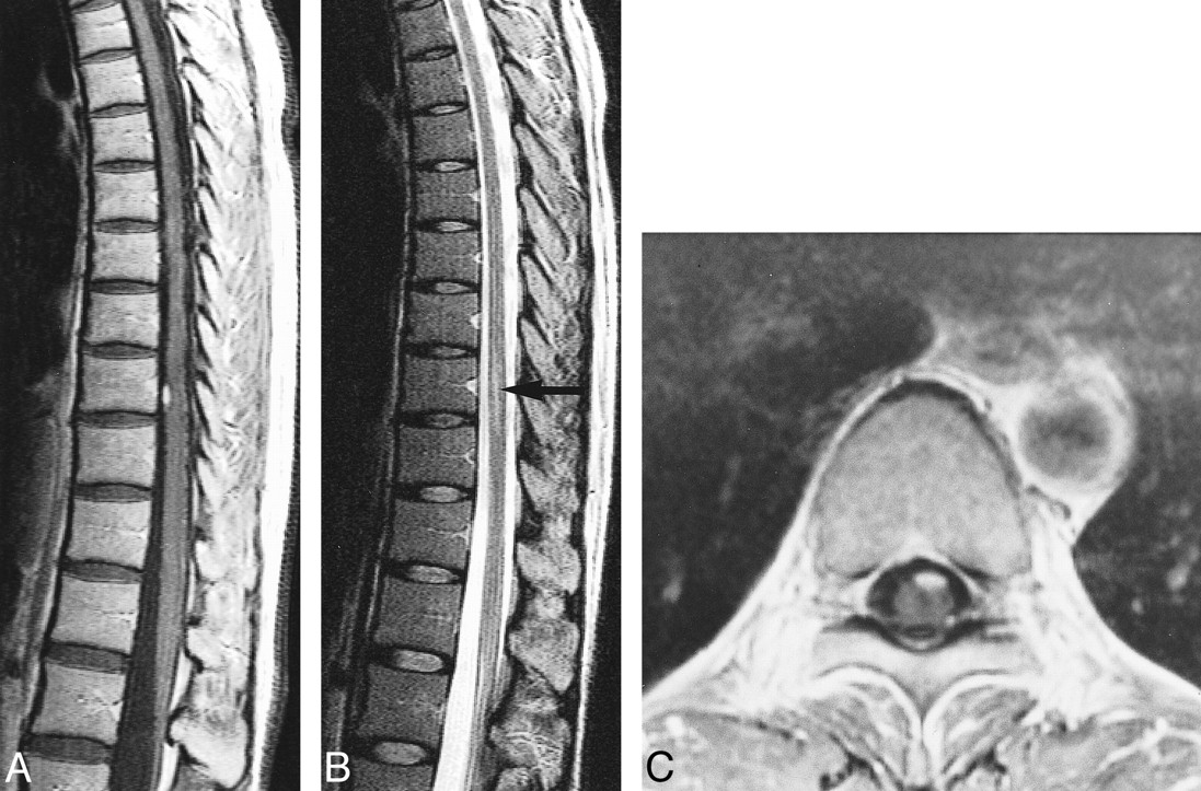
  • fig 6.
    • Download figure
    • Open in new tab
    • Download powerpoint
    fig 6.

    Case 1: 16-year-old boy with VHLD.

    A, Contrast-enhanced sagittal T1-weighted TSE image (700/12/2) shows a large, well-demarcated, intensely and heterogeneously enhancing intramedullary tumor at C7–T2. Note vascular flow voids in and around the tumor (curved arrows) and superficial enhancement of the spinal cord (straight arrows), proved to be dilated perimedullary veins at surgery. Syrinx extends from medulla oblongata to T12. Also note multiple small intensely and homogeneously enhancing tumors, most of which are located superficially at the posterior aspect of the spinal cord.

    B, Contrast-enhanced axial T1-weighted SE image (660/15/1) at C7–T1 shows enlargement of the spinal canal and the enhancing tumor. The tumor occupies entire dural sac and the spinal cord is barely discernible. The tumor was interpreted as deeply located within the spinal cord on MR image, and was confirmed at surgery.

  • fig 7.
    • Download figure
    • Open in new tab
    • Download powerpoint
    fig 7.

    Case 7: 49-year-old woman without VHLD.

    A and B, Noncontrast (A) and contrast-enhanced (B) sagittal T1-weighted SE images (600/15/2) show a small, homogeneously and intensely enhancing nodular tumor at C5–C6. The tumor is isointense on noncontrast T1-weighted image. Note extensive syrinx from medulla oblongata to T3 level.

    C, Contrast-enhanced axial T1-weighted SE image (600/15/2) shows the tumor is well demarcated, superficially located at the anterior aspect of the spinal cord.

  • fig 8.
    • Download figure
    • Open in new tab
    • Download powerpoint
    fig 8.

    Case 5: 49-year-old man with VHLD.

    A, Contrast-enhanced sagittal T1-weighted TSE image (700/12/3) shows a medium-sized, ovoid, heterogeneously enhancing tumor at T9. Note several small, homogeneously enhancing nodules cephalic to this tumor. Also note flow voids within the tumor and superficial enhancement of the spinal cord.

    B, Contrast-enhanced axial T1-weighted SE image (660/15/1) shows the tumor at T9 occupies entire dural sac. The tumor was thought to be located deeply within the spinal cord, but turned out to be subpial in location at surgery.

Tables

  • Figures
  • Table1

    Summary of clinical manifestations

PreviousNext
Back to top

In this issue

American Journal of Neuroradiology
Vol. 22, Issue 1
1 Jan 2001
  • Table of Contents
  • Index by author
Advertisement
Print
Download PDF
Email Article
Cite this article
0 Responses
Respond to this article
Share
Bookmark this article
MR Findings in Spinal Hemangioblastoma: Correlation with Symptoms and with Angiographic and Surgical Findings
Bao-Cheng Chu, Satoshi Terae, Kazutoshi Hida, Matakazu Furukawa, Satoru Abe, Kazuo Miyasaka
American Journal of Neuroradiology Jan 2001, 22 (1) 206-217;
del.icio.us logo Twitter logo Facebook logo Mendeley logo
  • Tweet Widget

Jump to section

  • Article
    • Abstract
    • Methods
    • Results
    • Discussion
    • Conclusion
    • Footnotes
    • References
  • Figures & Data
  • Info & Metrics
  • Responses
  • References
  • PDF

Related Articles

  • No related articles found.
  • PubMed
  • Google Scholar

Cited By...

  • Differentiating Hemangioblastomas from Brain Metastases Using Diffusion-Weighted Imaging and Dynamic Susceptibility Contrast-Enhanced Perfusion-Weighted MR Imaging
  • Magnetic Resonance Imaging of Spine Tumors: Classification, Differential Diagnosis, and Spectrum of Disease
  • Crossref
  • Google Scholar

This article has not yet been cited by articles in journals that are participating in Crossref Cited-by Linking.

More in this TOC Section

  • Clinical SVR of Fetal Brain MRI
  • FRACTURE MR in Congenital Vertebral Anomalies
  • Comparing MRI Perfusion in Pediatric Brain Tumors
Show more Pediatric Neuroimaging

Similar Articles

Advertisement

Indexed Content

  • Current Issue
  • Accepted Manuscripts
  • Article Preview
  • Past Issues
  • Editorials
  • Editor's Choice
  • Fellows' Journal Club
  • Letters to the Editor
  • Video Articles

Cases

  • Case Collection
  • Archive - Case of the Week
  • Archive - Case of the Month
  • Archive - Classic Case

More from AJNR

  • Trainee Corner
  • Imaging Protocols
  • MRI Safety Corner
  • Book Reviews

Multimedia

  • AJNR Podcasts
  • AJNR Scantastics

Resources

  • Turnaround Time
  • Submit a Manuscript
  • Submit a Video Article
  • Submit an eLetter to the Editor/Response
  • Manuscript Submission Guidelines
  • Statistical Tips
  • Fast Publishing of Accepted Manuscripts
  • Graphical Abstract Preparation
  • Imaging Protocol Submission
  • Evidence-Based Medicine Level Guide
  • Publishing Checklists
  • Author Policies
  • Become a Reviewer/Academy of Reviewers
  • News and Updates

About Us

  • About AJNR
  • Editorial Board
  • Editorial Board Alumni
  • Alerts
  • Permissions
  • Not an AJNR Subscriber? Join Now
  • Advertise with Us
  • Librarian Resources
  • Feedback
  • Terms and Conditions
  • AJNR Editorial Board Alumni

American Society of Neuroradiology

  • Not an ASNR Member? Join Now

© 2025 by the American Society of Neuroradiology All rights, including for text and data mining, AI training, and similar technologies, are reserved.
Print ISSN: 0195-6108 Online ISSN: 1936-959X

Powered by HighWire
Email this Article

Thank you for your interest in spreading the word on American Journal of Neuroradiology.

NOTE: We only request your email address so that the person you are recommending the page to knows that you wanted them to see it, and that it is not junk mail. We do not capture any email address.

Enter multiple addresses on separate lines or separate them with commas.
MR Findings in Spinal Hemangioblastoma: Correlation with Symptoms and with Angiographic and Surgical Findings
(Your Name) has sent you a message from American Journal of Neuroradiology
(Your Name) thought you would like to see the American Journal of Neuroradiology web site.
CAPTCHA
This question is for testing whether or not you are a human visitor and to prevent automated spam submissions.
Cite this article
Bao-Cheng Chu, Satoshi Terae, Kazutoshi Hida, Matakazu Furukawa, Satoru Abe, Kazuo Miyasaka
MR Findings in Spinal Hemangioblastoma: Correlation with Symptoms and with Angiographic and Surgical Findings
American Journal of Neuroradiology Jan 2001, 22 (1) 206-217;

Citation Manager Formats

  • BibTeX
  • Bookends
  • EasyBib
  • EndNote (tagged)
  • EndNote 8 (xml)
  • Medlars
  • Mendeley
  • Papers
  • RefWorks Tagged
  • Ref Manager
  • RIS
  • Zotero

We use cookies on this site to enhance your user experience. By clicking any link on this page you are giving your consent for us to set cookies.