Advertisement

Main menu

  • Home
  • Content
    • Current Issue
    • Accepted Manuscripts
    • Article Preview
    • Past Issue Archive
    • Video Articles
    • AJNR Case Collection
    • Case of the Week Archive
    • Case of the Month Archive
    • Classic Case Archive
  • Special Collections
    • AJNR Awards
    • Low-Field MRI
    • Alzheimer Disease
    • ASNR Foundation Special Collection
    • Photon-Counting CT
    • View All
  • Multimedia
    • AJNR Podcasts
    • AJNR SCANtastic
    • Trainee Corner
    • MRI Safety Corner
    • Imaging Protocols
  • For Authors
    • Submit a Manuscript
    • Submit a Video Article
    • Submit an eLetter to the Editor/Response
    • Manuscript Submission Guidelines
    • Statistical Tips
    • Fast Publishing of Accepted Manuscripts
    • Graphical Abstract Preparation
    • Imaging Protocol Submission
    • Author Policies
  • About Us
    • About AJNR
    • Editorial Board
    • Editorial Board Alumni
  • More
    • Become a Reviewer/Academy of Reviewers
    • Subscribers
    • Permissions
    • Alerts
    • Feedback
    • Advertisers
    • ASNR Home

User menu

  • Alerts
  • Log in

Search

  • Advanced search
American Journal of Neuroradiology

American Journal of Neuroradiology

ASHNR American Society of Functional Neuroradiology ASHNR American Society of Pediatric Neuroradiology ASSR
  • Alerts
  • Log in

Advanced Search

  • Home
  • Content
    • Current Issue
    • Accepted Manuscripts
    • Article Preview
    • Past Issue Archive
    • Video Articles
    • AJNR Case Collection
    • Case of the Week Archive
    • Case of the Month Archive
    • Classic Case Archive
  • Special Collections
    • AJNR Awards
    • Low-Field MRI
    • Alzheimer Disease
    • ASNR Foundation Special Collection
    • Photon-Counting CT
    • View All
  • Multimedia
    • AJNR Podcasts
    • AJNR SCANtastic
    • Trainee Corner
    • MRI Safety Corner
    • Imaging Protocols
  • For Authors
    • Submit a Manuscript
    • Submit a Video Article
    • Submit an eLetter to the Editor/Response
    • Manuscript Submission Guidelines
    • Statistical Tips
    • Fast Publishing of Accepted Manuscripts
    • Graphical Abstract Preparation
    • Imaging Protocol Submission
    • Author Policies
  • About Us
    • About AJNR
    • Editorial Board
    • Editorial Board Alumni
  • More
    • Become a Reviewer/Academy of Reviewers
    • Subscribers
    • Permissions
    • Alerts
    • Feedback
    • Advertisers
    • ASNR Home
  • Follow AJNR on Twitter
  • Visit AJNR on Facebook
  • Follow AJNR on Instagram
  • Join AJNR on LinkedIn
  • RSS Feeds

AJNR Awards, New Junior Editors, and more. Read the latest AJNR updates

Research ArticleBRAIN

Corpus Callosal Signal Changes in Patients with Obstructive Hydrocephalus after Ventriculoperitoneal Shunting

John I. Lane, Patrick H. Luetmer and John L. Atkinson
American Journal of Neuroradiology January 2001, 22 (1) 158-162;
John I. Lane
  • Find this author on Google Scholar
  • Find this author on PubMed
  • Search for this author on this site
Patrick H. Luetmer
  • Find this author on Google Scholar
  • Find this author on PubMed
  • Search for this author on this site
John L. Atkinson
  • Find this author on Google Scholar
  • Find this author on PubMed
  • Search for this author on this site
  • Article
  • Figures & Data
  • Info & Metrics
  • Responses
  • References
  • PDF
Loading

Article Figures & Data

Figures

  • Tables
  • fig 1.
    • Download figure
    • Open in new tab
    • Download powerpoint
    fig 1.

    Iatrogenic callosal injury. Axial proton density–weighted (2200/20/1) image shows small horizontal cleft of increased signal within the body of the corpus callosum (arrow) adjacent to the shunt catheter that is probably related to direct trauma sustained during catheter placement

  • fig 2.
    • Download figure
    • Open in new tab
    • Download powerpoint
    fig 2.

    Case 7: callosal injury from long-standing obstructive hydrocephalus.

    A, Sagittal T1-weighted (500/15/2) image obtained before shunt placement shows marked upward displacement of the corpus callosum (straight arrows). Note fourth ventricular mass (curved arrow) extending into and obstructing the aqueduct.

    B, Sagittal T1-weighted (500/20/2) image obtained 3 months after ventricular shunting shows diffuse areas of decreased signal within the body of the corpus callosum (arrows), which is likely the result of compression against the falx.

    C and D, Axial proton density– (C) and T2-weighted (D) (2200/20,80/1) images show increased signal within the body of the corpus callosum (arrows), corresponding to abnormalities in the sagittal plane.

  • fig 3.
    • Download figure
    • Open in new tab
    • Download powerpoint
    fig 3.

    Case 2: callosal injury from long-standing obstructive hydrocephalus in a patient with aqueductal stenosis.

    A, Sagittal T1-weighted (500/15/2) image obtained before shunt placement shows moderate upward displacement of the corpus callosum (arrows).

    B, T1-weighted sagittal (500/15/2) image 5 months after ventricular shunting shows diffuse areas of decreased signal within the posterior body of the corpus callosum (arrows), which was most likely caused by compression against the falx.

    C and D, Axial proton density– and T2-weighted (2200/20,80/1) images show increased signal within the body of the corpus callosum (arrows) corresponding to abnormalities in the sagittal plane.

  • fig 4.
    • Download figure
    • Open in new tab
    • Download powerpoint
    fig 4.

    Case 1: callosal injury in a patient with obstructing tectal tumor. Postshunt sagittal FLAIR sequence (11002/148/1, TI = 2250) proved to be the most sensitive in detecting callosal signal changes but was not included in the imaging protocols of all examinations reviewed

Tables

  • Figures
  • Table1

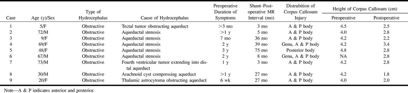
    Subgroup of shunted patients with corpus callosal signal changes

PreviousNext
Back to top

In this issue

American Journal of Neuroradiology
Vol. 22, Issue 1
1 Jan 2001
  • Table of Contents
  • Index by author
Advertisement
Print
Download PDF
Email Article

Thank you for your interest in spreading the word on American Journal of Neuroradiology.

NOTE: We only request your email address so that the person you are recommending the page to knows that you wanted them to see it, and that it is not junk mail. We do not capture any email address.

Enter multiple addresses on separate lines or separate them with commas.
Corpus Callosal Signal Changes in Patients with Obstructive Hydrocephalus after Ventriculoperitoneal Shunting
(Your Name) has sent you a message from American Journal of Neuroradiology
(Your Name) thought you would like to see the American Journal of Neuroradiology web site.
CAPTCHA
This question is for testing whether or not you are a human visitor and to prevent automated spam submissions.
Cite this article
John I. Lane, Patrick H. Luetmer, John L. Atkinson
Corpus Callosal Signal Changes in Patients with Obstructive Hydrocephalus after Ventriculoperitoneal Shunting
American Journal of Neuroradiology Jan 2001, 22 (1) 158-162;

Citation Manager Formats

  • BibTeX
  • Bookends
  • EasyBib
  • EndNote (tagged)
  • EndNote 8 (xml)
  • Medlars
  • Mendeley
  • Papers
  • RefWorks Tagged
  • Ref Manager
  • RIS
  • Zotero
0 Responses
Respond to this article
Share
Bookmark this article
Corpus Callosal Signal Changes in Patients with Obstructive Hydrocephalus after Ventriculoperitoneal Shunting
John I. Lane, Patrick H. Luetmer, John L. Atkinson
American Journal of Neuroradiology Jan 2001, 22 (1) 158-162;
del.icio.us logo Twitter logo Facebook logo Mendeley logo
  • Tweet Widget
  • Facebook Like
  • Google Plus One

Jump to section

  • Article
    • Abstract
    • Methods
    • Results
    • Discussion
    • Conclusion
    • Footnotes
    • References
  • Figures & Data
  • Info & Metrics
  • Responses
  • References
  • PDF

Related Articles

  • No related articles found.
  • PubMed
  • Google Scholar

Cited By...

  • Corpus callosum impingement syndrome
  • Corpus Callosum Hyperintensity in Normal Pressure Hydrocephalus After Ventriculoperitoneal Shunt
  • Signal hyperintensity of the callosum after ventriculoperitoneal shunting
  • Functional and magnetic resonance imaging correlates of corpus callosum in normal pressure hydrocephalus before and after shunting
  • Crossref
  • Google Scholar

This article has not yet been cited by articles in journals that are participating in Crossref Cited-by Linking.

More in this TOC Section

  • Multimodal CT Provides Improved Performance for Lacunar Infarct Detection
  • Optimal MRI Sequence for Identifying Occlusion Location in Acute Stroke: Which Value of Time-Resolved Contrast-Enhanced MRA?
  • Evaluating the Effects of White Matter Multiple Sclerosis Lesions on the Volume Estimation of 6 Brain Tissue Segmentation Methods
Show more BRAIN

Similar Articles

Advertisement

Indexed Content

  • Current Issue
  • Accepted Manuscripts
  • Article Preview
  • Past Issues
  • Editorials
  • Editor's Choice
  • Fellows' Journal Club
  • Letters to the Editor
  • Video Articles

Cases

  • Case Collection
  • Archive - Case of the Week
  • Archive - Case of the Month
  • Archive - Classic Case

More from AJNR

  • Trainee Corner
  • Imaging Protocols
  • MRI Safety Corner
  • Book Reviews

Multimedia

  • AJNR Podcasts
  • AJNR Scantastics

Resources

  • Turnaround Time
  • Submit a Manuscript
  • Submit a Video Article
  • Submit an eLetter to the Editor/Response
  • Manuscript Submission Guidelines
  • Statistical Tips
  • Fast Publishing of Accepted Manuscripts
  • Graphical Abstract Preparation
  • Imaging Protocol Submission
  • Evidence-Based Medicine Level Guide
  • Publishing Checklists
  • Author Policies
  • Become a Reviewer/Academy of Reviewers
  • News and Updates

About Us

  • About AJNR
  • Editorial Board
  • Editorial Board Alumni
  • Alerts
  • Permissions
  • Not an AJNR Subscriber? Join Now
  • Advertise with Us
  • Librarian Resources
  • Feedback
  • Terms and Conditions
  • AJNR Editorial Board Alumni

American Society of Neuroradiology

  • Not an ASNR Member? Join Now

© 2025 by the American Society of Neuroradiology All rights, including for text and data mining, AI training, and similar technologies, are reserved.
Print ISSN: 0195-6108 Online ISSN: 1936-959X

Powered by HighWire