
Article Figures & Data
Figures
- fig 1.
Case 1: Eight-year-old girl with crouzonoid features, acanthosis nigricans, and FGFR3 ala391glu mutation.
A, Helical CT shows bilateral JFA (arrows) and a large left mastoid emissary foramen (arrowheads).
B, Axial collapsed maximum intensity projection (MIP) from 3D TOF MRV (53/6.9/1 [TR/TE/excitations]) reveals atresia of the right transverse and sigmoid sinuses and the both internal JV (arrowheads). Large occipitomastoid EV arise from the left transverse and sigmoid sinuses (arrows).
- fig 2.
Case 3: Three-year-old boy with crouzonoid features, acanthosis nigricans, and FGFR3 ala391glu mutation.
A, Axial CT shows a large central occipital emissary foramen (arrow). A small left mastoid emissary foramen is seen (arrowhead).
B, Axial CT 2 years after surgical clipping of the occipital emissary vein (arrowhead) shows marked enlargement of the left mastoid emissary foramen (arrow).
C, Digital subtraction cerebral angiogram, frontal projection, shows atresia of the right sigmoid sinus and lateral portion of the left sigmoid sinus (arrows) and atresia of both internal JV. There are large left mastoid EV arising from the proximal left sigmoid sinus (arrowheads).
- fig 3.
Case 5: Three-year-old girl with Pfeiffer syndrome and FGFR2 cys342arg mutation. Frontal MIP of a CT venogram shows occlusion of the transverse sinuses (arrowheads) and large duplicated occipital sinuses (arrows). fig 4. Case 20: Ten-year-old girl with Pfeiffer syndrome and FGFR2 ser354cys mutation. Helical CT shows a large right occipitomastoid emissary foramen (arrowhead). The right jugular foramen is stenotic, with prominent septations (arrow). The left JF appeared stenotic on a caudal image of the skull base (not shown)
- fig 5.
Case 12: A 21-month-old girl with Crouzon syndrome and FGFR2 cys342tyr mutation. Axial collapsed MIP from 3D TOF MRV (53/6.9/1) 20 months after the initial CT shows JFS. There is stenosis of both transverse and sigmoid sinuses and the internal JV (arrowheads). There are large, right, occipitomastoid EV (arrows). fig 6. Case 23: Three-year-old boy with clinically unclassified, bilateral, coronal synostosis and FGFR2 ala314ser mutation. Axial T1-weighted MR imaging (450/14/1) reveals flow voids within large bilateral mastoid EV (arrows). The JV are stenotic (arrowheads)
- fig 7.
Case 24: Two-year-old girl with Crouzon syndrome and FGFR ser354cys mutation.
A, Axial CT shows that the JF are not stenotic (asterisks).
B, Magnified, oblique, frontal MIP from 3D TOF MRV (53/6.9/1) reveals a focal stenosis at the junction of the right sigmoid sinus with the internal JV (arrow). There is a large right mastoid EV (arrowheads).


Tables
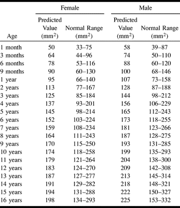
 TABLE 1:
Predicted value and normal ranges for the sum of the cross-sectional area of the right and left jugular foramina according to age and gender
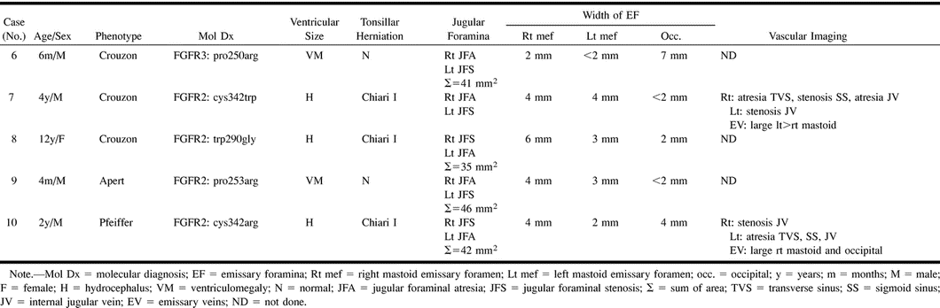
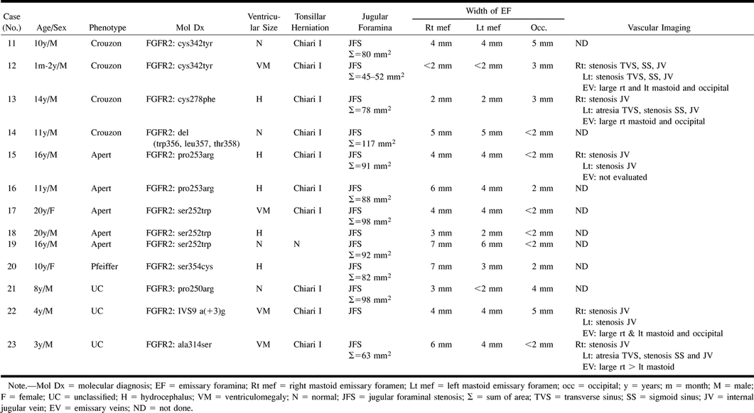
- TABLE 3:
Group 2. Unilateral jugular foraminal atresia and contralateral jugular foraminal stenosis
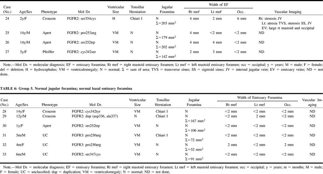
- TABLE 5:
Group 4. Normal jugular foramina; prominent basal emissary foramina
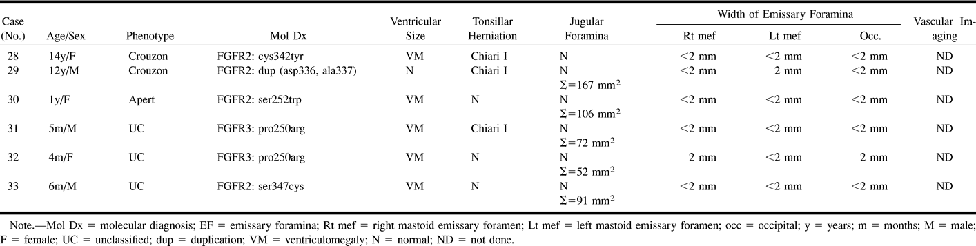
- TABLE 6:
Group 5. Normal jugular foramina; normal basal emissary foramina
In this issue


Jump to section
Related Articles
Cited By...
- No citing articles found.