Advertisement

Main menu

  • Home
  • Content
    • Current Issue
    • Accepted Manuscripts
    • Article Preview
    • Past Issue Archive
    • Video Articles
    • AJNR Case Collection
    • Case of the Week Archive
    • Case of the Month Archive
    • Classic Case Archive
  • Special Collections
    • AJNR Awards
    • Low-Field MRI
    • Alzheimer Disease
    • ASNR Foundation Special Collection
    • Photon-Counting CT
    • View All
  • Multimedia
    • AJNR Podcasts
    • AJNR SCANtastic
    • Trainee Corner
    • MRI Safety Corner
    • Imaging Protocols
  • For Authors
    • Submit a Manuscript
    • Submit a Video Article
    • Submit an eLetter to the Editor/Response
    • Manuscript Submission Guidelines
    • Statistical Tips
    • Fast Publishing of Accepted Manuscripts
    • Graphical Abstract Preparation
    • Imaging Protocol Submission
    • Author Policies
  • About Us
    • About AJNR
    • Editorial Board
    • Editorial Board Alumni
  • More
    • Become a Reviewer/Academy of Reviewers
    • Subscribers
    • Permissions
    • Alerts
    • Feedback
    • Advertisers
    • ASNR Home

User menu

  • Alerts
  • Log in

Search

  • Advanced search
American Journal of Neuroradiology

American Journal of Neuroradiology

ASHNR American Society of Functional Neuroradiology ASHNR American Society of Pediatric Neuroradiology ASSR
  • Alerts
  • Log in

Advanced Search

  • Home
  • Content
    • Current Issue
    • Accepted Manuscripts
    • Article Preview
    • Past Issue Archive
    • Video Articles
    • AJNR Case Collection
    • Case of the Week Archive
    • Case of the Month Archive
    • Classic Case Archive
  • Special Collections
    • AJNR Awards
    • Low-Field MRI
    • Alzheimer Disease
    • ASNR Foundation Special Collection
    • Photon-Counting CT
    • View All
  • Multimedia
    • AJNR Podcasts
    • AJNR SCANtastic
    • Trainee Corner
    • MRI Safety Corner
    • Imaging Protocols
  • For Authors
    • Submit a Manuscript
    • Submit a Video Article
    • Submit an eLetter to the Editor/Response
    • Manuscript Submission Guidelines
    • Statistical Tips
    • Fast Publishing of Accepted Manuscripts
    • Graphical Abstract Preparation
    • Imaging Protocol Submission
    • Author Policies
  • About Us
    • About AJNR
    • Editorial Board
    • Editorial Board Alumni
  • More
    • Become a Reviewer/Academy of Reviewers
    • Subscribers
    • Permissions
    • Alerts
    • Feedback
    • Advertisers
    • ASNR Home
  • Follow AJNR on Twitter
  • Visit AJNR on Facebook
  • Follow AJNR on Instagram
  • Join AJNR on LinkedIn
  • RSS Feeds

AJNR Awards, New Junior Editors, and more. Read the latest AJNR updates

Research ArticlePediatric Neuroimaging

MR, CT, and Plain Film Imaging of the Developing Skull Base in Fetal Specimens

William R. Nemzek, Hilary A. Brodie, Stephen T. Hecht, Brian W. Chong, Catherine J. Babcook and J. Anthony Seibert
American Journal of Neuroradiology October 2000, 21 (9) 1699-1706;
William R. Nemzek
  • Find this author on Google Scholar
  • Find this author on PubMed
  • Search for this author on this site
Hilary A. Brodie
  • Find this author on Google Scholar
  • Find this author on PubMed
  • Search for this author on this site
Stephen T. Hecht
  • Find this author on Google Scholar
  • Find this author on PubMed
  • Search for this author on this site
Brian W. Chong
  • Find this author on Google Scholar
  • Find this author on PubMed
  • Search for this author on this site
Catherine J. Babcook
  • Find this author on Google Scholar
  • Find this author on PubMed
  • Search for this author on this site
J. Anthony Seibert
  • Find this author on Google Scholar
  • Find this author on PubMed
  • Search for this author on this site
  • Article
  • Figures & Data
  • Info & Metrics
  • Responses
  • References
  • PDF
Loading

Article Figures & Data

Figures

  • Tables
  • Figure1
    • Download figure
    • Open in new tab
    • Download powerpoint

    fig 1. T2-weighted fast spin-echo MR image (4000/90/4) of fetal specimen with a gestational age of 11 weeks 5 days shows that the skull base is formed in cartilage before ossification. The basiocciput (straight solid arrow), the postsphenoid (open arrow), containing the pituitary in the cartilaginous sella turcica, and the presphenoid (curved arrow) are seen. Note the optic chiasm (arrowhead). The cerebral hemispheres and anterior third ventricle are derived from the telencephalon. The cerebral hemispheres consist of lateral diverticula that have not yet expanded to cover the brain stem. Note ossification of palate and mandible. fig 2. T2-weighted fast spin-echo MR image (500/90/3) of a fetal specimen with a gestational age of 14 weeks 4 days shows ossification as low signal in the supraoccipital and basioccipital (short straight arrow) centers of the occipital bone. The cartilaginous sella turcica contains the pituitary (open arrow). Note the optic chiasm superior to the chiasmatic sulcus (curved arrow) of the presphenoid. The chiasmatic sulcus is delineated anteriorly by the limbus and posteriorly by the tuberculum sellae (ts)

  • fig 3.
    • Download figure
    • Open in new tab
    • Download powerpoint
    fig 3.

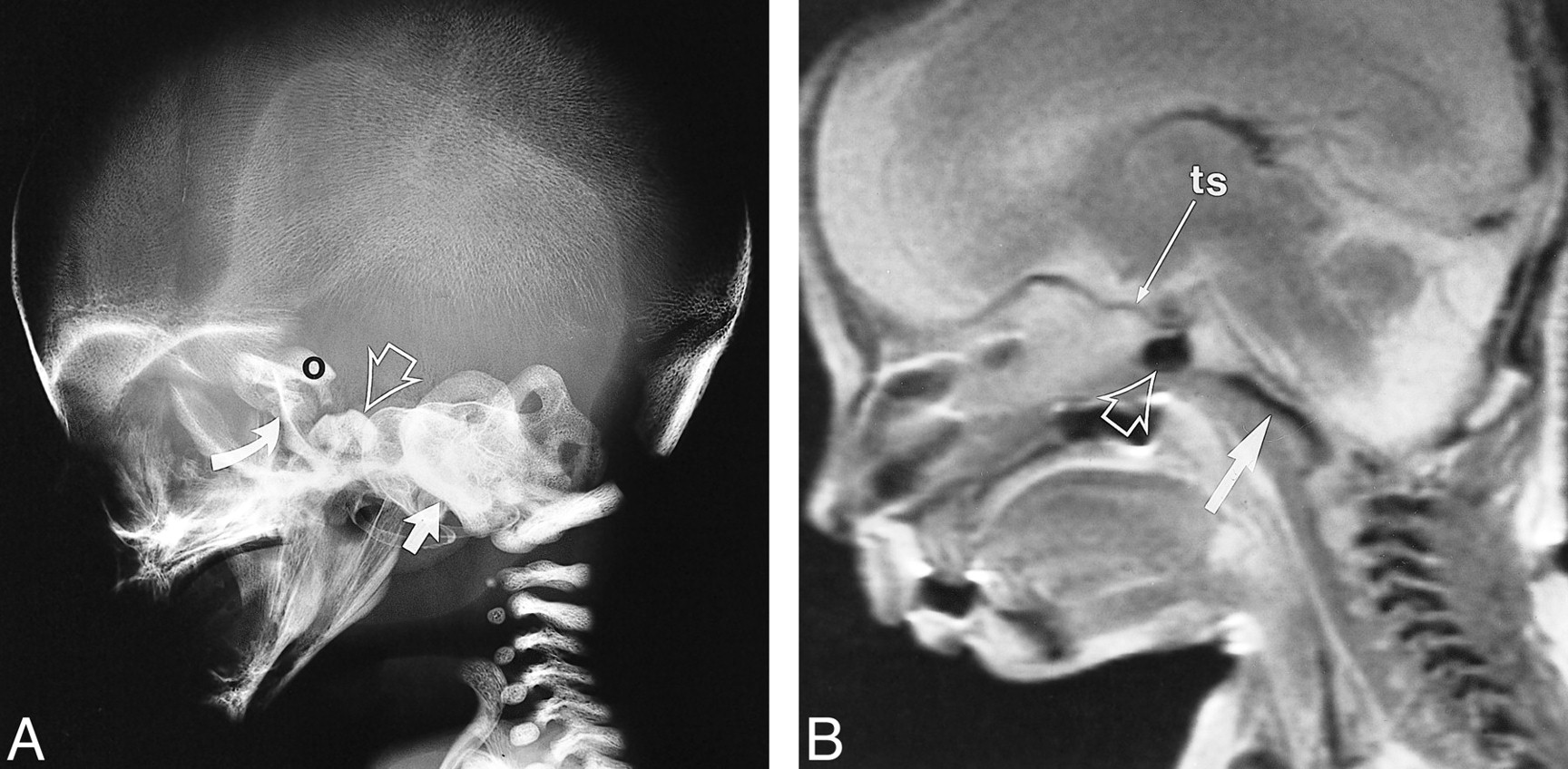
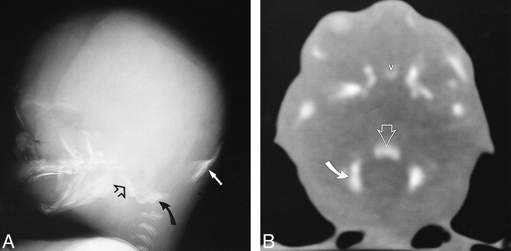
    A and B, Lateral skull radiograph of a fetal specimen with a gestational age of 19 weeks 5 days (A) and T2-weighted fast spin-echo MR image (4500/90/4) of a fetal specimen with a gestational age of 19 weeks 3 days (B) show progressive ossification of the skull base from posterior to anterior. The ossification has extended to the postsphenoid (open arrow), which encloses the pituitary to form the sella turcica. The cerebral hemispheres have greatly expanded. Note ossification of presphenoid (curved arrow) on lateral radiograph (arrow, basiocciput; O, orbitosphenoid; ts, tuberculum sellae)

  • fig 3.
    • Download figure
    • Open in new tab
    • Download powerpoint
    fig 3.

    fig 4. T2-weighted fast spin-echo MR image (4500/87/4) of a fetal specimen with a gestational age of 20 weeks 5 days shows progressive skull base ossification that has extended from the basiocciput (straight arrow) to the postsphenoid (open arrow) to the presphenoid (curved arrow) in the area of the chiasmatic sulcus. fig 5. A and B, Lateral skull radiograph of a fetal specimen with a gestational age of 22 weeks 3 days (A) and T2-weighted fast spin-echo MR image (4000/90/3) of a fetal specimen with a gestational age of 24 weeks 4 days (B) show advancing ossification of the basiocciput (arrow), basisphenoid (postsphenoid, open arrow), and presphenoid (curved arrow). Most of the growth of the head is due to the dominant development of the cerebral hemispheres (O, orbitosphenoid)

  • fig 6.
    • Download figure
    • Open in new tab
    • Download powerpoint
    fig 6.

    CT scans at 16 weeks 2 days, 18 weeks 4 days, 18 weeks 5 days, and 24 weeks 4 days show progressive ossification of the postsphenoid. Note that the medial ossification centers (solid arrows) appear first followed by ossification of the lateral centers (open arrows)

  • fig 6.
    • Download figure
    • Open in new tab
    • Download powerpoint
    fig 6.

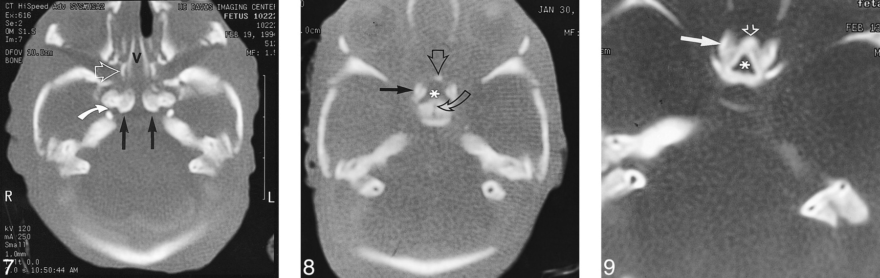
    fig 7. CT scan of fetal specimen with a gestational age of 23 weeks 3 days shows persistence of the medial ossification centers of the postsphenoid as separate structures (straight solid arrows). Note the bones of Bertin (open arrow) adjacent to the vomer (v) and rostrum of the basisphenoid. This is the site of sphenoid pneumatization. Note also carotid groove in sphenoid (curved arrow). fig 8. CT scan of fetal specimen with a gestational age of 21 weeks 4 days shows early ossification of the main centers (solid arrow) and corporal middle centers (open straight arrow) of the presphenoid. Note small defect that represents the remnant of the obliterated craniopharyngeal canal (Rathke's pouch) in the postsphenoid (curved arrow). Asterisk indicates olivary eminence. fig 9. CT scan of fetal specimen with a gestational age of 24 weeks 4 days shows progressive ossification and fusion of the main (solid arrow) and corporal middle (open arrow) portions of the presphenoid. Note the olivary eminence (asterisk), which is a triangular area filled with unossified cartilage in the presphenoid

  • fig 6.
    • Download figure
    • Open in new tab
    • Download powerpoint
    fig 6.

    fig 10. CT scans show progressive ossification of the orbitosphenoid (arrow) at 16 weeks 2 days, 17 weeks 4 days, 18 weeks 4 days, and 24 weeks 4 days. fig 11. CT scan of fetal specimen with a gestational age of 22 weeks 3 days shows ossification of alisphenoid (A), which forms the greater wing of the sphenoid. Foramen ovale and spinosum are seen as a large defect in the greater wing of the sphenoid (arrow)

  • fig 12.
    • Download figure
    • Open in new tab
    • Download powerpoint
    fig 12.

    Diagram of the skull base ossification centers. Asterisk indicates olivary eminence (anterior foramen)

  • fig 13.
    • Download figure
    • Open in new tab
    • Download powerpoint
    fig 13.

    Diagram of progression of skull base ossification. Arrow indicates normal sequence of ossification of the skull base from posterior to anterior

  • fig 14.
    • Download figure
    • Open in new tab
    • Download powerpoint
    fig 14.

    A and B, Lateral radiograph (A) and CT scan (B) of fetal specimen with a gestational age of 16 weeks 2 days show early ossification of the four primary centers of the occipital bone that surrounds the foramen magnum: supraoccipital (solid arrow), basioccipital (open arrows), and exoccipital (curved arrows). Note ossification of the frontal bone, vomer (v), pterygoid plates, zygoma, mandible, and maxilla

Tables

  • Figures
  • TABLE 1:
    • View popup
    • Download powerpoint
    TABLE 1:

    Skull base ossification

  • TABLE 2:
    • View popup
    • Download powerpoint
    TABLE 2:

    Milestones in skull base development and corresponding imaging findings

PreviousNext
Back to top

In this issue

American Journal of Neuroradiology
Vol. 21, Issue 9
1 Oct 2000
  • Table of Contents
  • Index by author
Advertisement
Print
Download PDF
Email Article

Thank you for your interest in spreading the word on American Journal of Neuroradiology.

NOTE: We only request your email address so that the person you are recommending the page to knows that you wanted them to see it, and that it is not junk mail. We do not capture any email address.

Enter multiple addresses on separate lines or separate them with commas.
MR, CT, and Plain Film Imaging of the Developing Skull Base in Fetal Specimens
(Your Name) has sent you a message from American Journal of Neuroradiology
(Your Name) thought you would like to see the American Journal of Neuroradiology web site.
CAPTCHA
This question is for testing whether or not you are a human visitor and to prevent automated spam submissions.
Cite this article
William R. Nemzek, Hilary A. Brodie, Stephen T. Hecht, Brian W. Chong, Catherine J. Babcook, J. Anthony Seibert
MR, CT, and Plain Film Imaging of the Developing Skull Base in Fetal Specimens
American Journal of Neuroradiology Oct 2000, 21 (9) 1699-1706;

Citation Manager Formats

  • BibTeX
  • Bookends
  • EasyBib
  • EndNote (tagged)
  • EndNote 8 (xml)
  • Medlars
  • Mendeley
  • Papers
  • RefWorks Tagged
  • Ref Manager
  • RIS
  • Zotero
0 Responses
Respond to this article
Share
Bookmark this article
MR, CT, and Plain Film Imaging of the Developing Skull Base in Fetal Specimens
William R. Nemzek, Hilary A. Brodie, Stephen T. Hecht, Brian W. Chong, Catherine J. Babcook, J. Anthony Seibert
American Journal of Neuroradiology Oct 2000, 21 (9) 1699-1706;
del.icio.us logo Twitter logo Facebook logo Mendeley logo
  • Tweet Widget
  • Facebook Like
  • Google Plus One

Jump to section

  • Article
    • Abstract
    • Methods
    • Results
    • Discussion
    • Conclusion
    • Acknowledgments
    • Footnotes
    • References
  • Figures & Data
  • Info & Metrics
  • Responses
  • References
  • PDF

Related Articles

  • No related articles found.
  • PubMed
  • Google Scholar

Cited By...

  • Sphenoid Bone Pneumatisation on Lateral Cephalograms of Patients With Neurofibromatosis Type 1
  • Characteristic MR Imaging Findings of the Neonatal Brain in RASopathies
  • MR Imaging of the Pituitary Gland and Postsphenoid Ossification in Fetal Specimens
  • Crossref
  • Google Scholar

This article has not yet been cited by articles in journals that are participating in Crossref Cited-by Linking.

More in this TOC Section

  • Neuroimaging Delineation and Progression of SLSMD
  • fetal brain development of 10 weeks gestation
  • CHARGE fetal MRI clival cleft
Show more Pediatric Neuroimaging

Similar Articles

Advertisement

Indexed Content

  • Current Issue
  • Accepted Manuscripts
  • Article Preview
  • Past Issues
  • Editorials
  • Editor's Choice
  • Fellows' Journal Club
  • Letters to the Editor
  • Video Articles

Cases

  • Case Collection
  • Archive - Case of the Week
  • Archive - Case of the Month
  • Archive - Classic Case

More from AJNR

  • Trainee Corner
  • Imaging Protocols
  • MRI Safety Corner
  • Book Reviews

Multimedia

  • AJNR Podcasts
  • AJNR Scantastics

Resources

  • Turnaround Time
  • Submit a Manuscript
  • Submit a Video Article
  • Submit an eLetter to the Editor/Response
  • Manuscript Submission Guidelines
  • Statistical Tips
  • Fast Publishing of Accepted Manuscripts
  • Graphical Abstract Preparation
  • Imaging Protocol Submission
  • Evidence-Based Medicine Level Guide
  • Publishing Checklists
  • Author Policies
  • Become a Reviewer/Academy of Reviewers
  • News and Updates

About Us

  • About AJNR
  • Editorial Board
  • Editorial Board Alumni
  • Alerts
  • Permissions
  • Not an AJNR Subscriber? Join Now
  • Advertise with Us
  • Librarian Resources
  • Feedback
  • Terms and Conditions
  • AJNR Editorial Board Alumni

American Society of Neuroradiology

  • Not an ASNR Member? Join Now

© 2025 by the American Society of Neuroradiology All rights, including for text and data mining, AI training, and similar technologies, are reserved.
Print ISSN: 0195-6108 Online ISSN: 1936-959X

Powered by HighWire