
Article Figures & Data
Figures
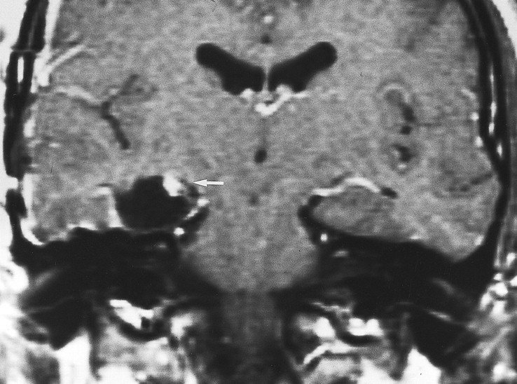
- fig 1.
Typical postoperative changes of the choroid plexus after temporal lobectomy. Coronal postcontrast T1-weighted MR image, obtained in the immediate postoperative period (1 day) after right temporal lobectomy, shows enlargement and intense nodular enhancement of the choroid plexus on the right (arrow) compared with the normal side
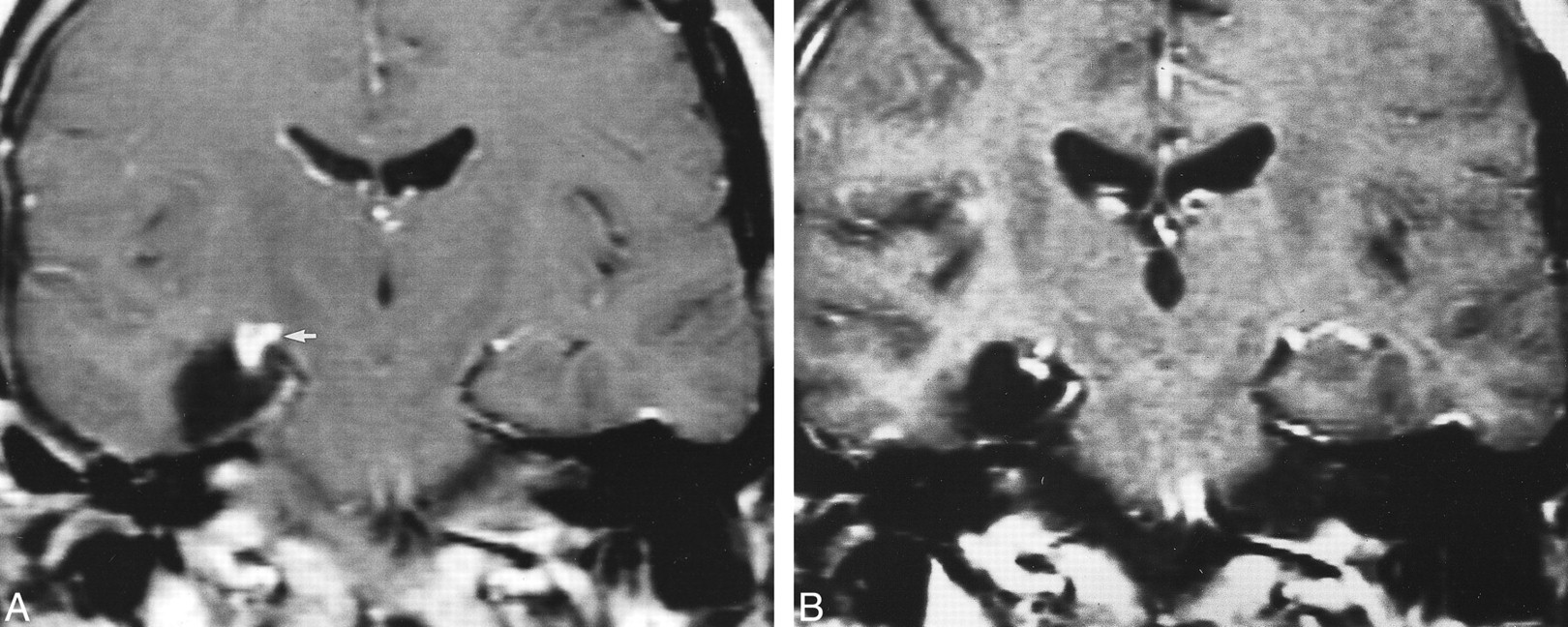
- fig 2.
Postoperative changes of choroid plexus with time, on enhanced T1-weighted coronal images. A, Intense, nodular enhancement of the choroid plexus (arrow) on the right side in the immediate (1 day) postoperative period. B, Persistent nodular changes 9 months after surgery, although the enlargement and the degree of enhancement have markedly diminished in the interval
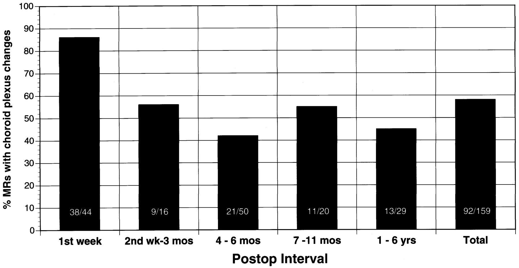
- fig 3.
This graph illustrates the frequency of the choroid plexus changes as a function of time. Thirty-eight (86%) of the 44 MR scans performed within the 1st week after surgery revealed an enlarged, intensely enhancing choroid plexus. These choroid plexus changes persist in about half the cases after the 1st postoperative week
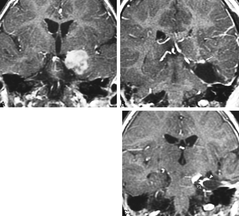
- fig 4.
Recurrent neoplastic disease with similar appearance to postoperative choroid plexus enhancement revealed on coronal enhanced T1-weighted images. A, The preoperative image shows a large, heterogeneously enhancing tumor involving the medial temporal lobe. B, MR scan 1 month after resective surgery shows a prolapsed choroid plexus adherent to the surgical margins (arrow). C, At 3 months after surgery, several enhancing foci are present. Recurrent enhancing oligodendroglioma was found medially (arrow), as confirmed on sequential images (not seen). The choroid plexus enhancement (arrowhead) adjacent to the tumor as well as the dural enhancement (open arrows) could be misinterpreted as neoplastic enhancement.
- fig 5.
Postsurgical choroid plexus herniation on coronal, enhanced, T1-weighted images. A, Preoperative scan shows the normal-appearing choroid plexus in the lateral ventricles. B, Immediate postoperative scan depicts herniation of the choroid plexus inferiorly into the surgical site (arrow). C, Persistent sagging of the choroid plexus 6 months (arrow) after surgery. This should be distinguished from adjacent postoperative dural enhancement (arrowhead).


