
Article Figures & Data
Figures
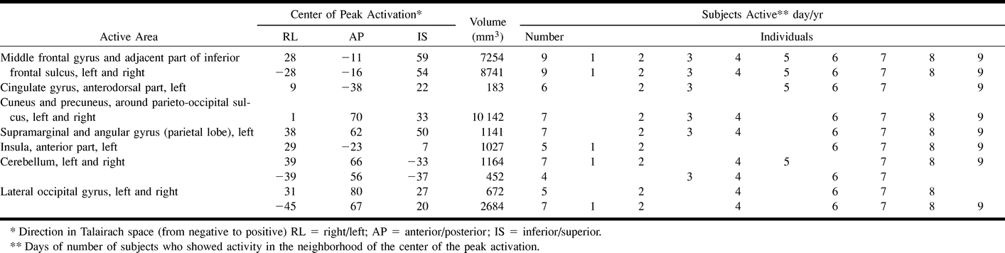
- fig 1.
Example of the Tower of London screen.
A, Sample screen of one of the configurations of a planning problem. Upper, baseline configuration; lower, target configuration. In this example, the participant has been asked to move first the blue ball to the right rod, which is counterintuitive. Thereafter, the participant has to place the yellow ball on top of the red ball, the blue ball at its destination, the yellow ball on top of the blue ball, the red ball on the right rod, and, finally, the yellow ball at the target position (sixth move). Two alternatives are presented on each side of the screen, from which the participant had to choose the correct answer. The participant was asked to respond by pressing the air bulb at the corresponding side.
B, Sample screen of the control configuration. The participant has to count the yellow and blue balls altogether. In this example, the answer is six, which is indicated on the right.
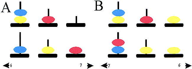
- fig 2.
Overview of the test, imaging, and data analysis. With this task paradigm, easy and difficult planning and counting are performed in blocks. Every block lasts 36 s, including a 4-s instruction. A total of nine blocks were performed during the test. A total of 82 images were obtained, including those of a dummy before the test started. To account for the hemodynamic response delay, a 4-s delay in the analysis was used. The images obtained during the instruction (accounting for the hemodynamic response delay) were not used for further calculations. The resulting 71 images were used for the analysis (24 obtained during the difficult planning problems, 24 obtained during the easy planning problems, and 23 obtained during the control stage)
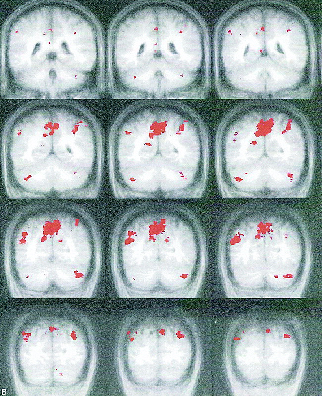
- fig 3.
Activated areas during the active condition of the Tower of London task. During the active condition (planning stage) of the task, activation (red) on the group average map (shown in Talairach format with coronal orientation) is shown. In the brain area from −3 anterior to +49 posterior, no activation was seen. A, Frontal regions. Coronal sections of coordinates −44 to −3 (anterior part of the brain). Activity was noted in the dorsolateral prefrontal cortex, the anterior part of the cingulate cortex, a part of the precentral cortex, and the frontal opercular area of the insula during planning. The right side shows slightly more activation than the left side. B, Parietal/occipital regions. Coronal sections of coordinates +49 to +80 (posterior part of the brain). Activation was noted in the cuneus and precuneus region, the marginal and angular gyrus in the parietal lobe, and the cerebellum during the active condition


Tables

Areas of fMR activation during the Tower of London task; planning condition (mean of all subjects)
In this issue


Jump to section
Related Articles
Cited By...
- Sleep spindles and slow waves are physiological markers for age-related changes in gray matter in brain regions supporting problem-solving skills
- Catechol O-Methyltransferase val158met Genotype Influences Frontoparietal Activity during Planning in Patients with Parkinson's Disease
- Health related quality of life in adults with repaired tetralogy of Fallot: psychosocial and cognitive outcomes