
Article Figures & Data
Figures
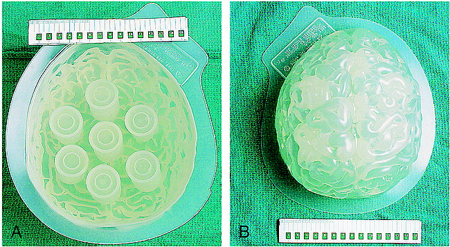
- fig 1.
Gel-filled brain phantom. A, Anterior view and B, posterior view
- fig 2.
T1-weighted (top) and functional MR (bottom) images of the brain phantom
- fig 3.
Example of six motion parameters (three translation and three rotation) as determined by the realignment algorithm in response to a step-function displacement. Note that movements are virtually all in-plane (translation in x and y, rotation about z), with translations and rotations limited to less than 1 mm and less than 1 degree, respectively. The highest correlation with the boxcar reference waveform (r = 0.52) is for rotation about z (in-plane). The abscissae are scaled in frames, with TR = 3 s/frame
- fig 4.
Numbers of falsely activated regions (at P < .05, corrected for multiple comparisons), excluding phantom edges and regions larger than 300 voxels, plotted against the maximum coefficient of correlation between motion and boxcar reference for each experiment. Each experiment is plotted twice, with a circle and triangle representing the low (P = .005) and high (P = .001) thresholds, respectively, applied to statistical parametric maps. There are 12 data points at the origin representing the six control experiments. Note that for correlations of r < 0.52, no areas of false activation were seen. For r > 0.67, false activation was observed in every experiment. Clusters became more coalescent at higher correlations; this explains the apparent lack of correlation between cluster number and r.
- fig 5.
Sizes of falsely activated regions (at P < .05, corrected for multiple comparisons), excluding phantom edges and regions larger than 300 voxels, plotted against the maximum coefficient of correlation between motion and boxcar reference for each experiment. There are six data points at the origin on each plot, representing the six control experiments.
A, Plots for low (P = .005) thresholds applied to statistical parametric maps.
B, Plots for high (P = .001) thresholds applied to statistical parametric maps.
- fig 6.
Activation maps overlaid onto functional MR images in sagittal (upper left), axial (lower left), and coronal (upper right) planes for an experiment with r = 0.70. Clusters were obtained from the statistical parametric map at a threshold of P = .001 (Z = 3.09). Crosshairs point to a 159-voxel region of false activation significant at P < .05, corrected for multiple comparisons. A second significant region of false activation is seen in the lower right on the coronal image. Several “activated” regions seen along the air-phantom edges were excluded from cluster counts (several of these smaller surface artifacts project over the center of the coronal image but do not lie within the middle of the phantom and are not counted). Functional MR imaging pulse sequence: 2D spiral; 3000/5; flip angle, 88°
- fig 7.
Activation map overlaid onto functional MR images in axial (top) and coronal (bottom) planes for a resting human volunteer subjected to passive, in-plane head motion (correlation of motion with boxcar reference, r = 0.75). Local activation is seen in left parietal cortex. Is this an area of motion-induced false activation? Functional MR imaging pulse sequence: 2D spiral; 3000/5; flip angle, 88°
- fig 8.
Functional MR imaging signal at a falsely activated voxel, after realignment and temporal smoothing, is shown along with the x-translation vector (same experiment as that shown in fig 3). Failure of the realignment algorithm to correct fully for motion effects is clearly evident. (The abscissa is scaled in frames, with TR = 3 s/frame. The MR signal intensity scale is arbitrary, with approximately 2% change from lowest to highest value.)
- fig 9.
Examples of theoretical head movements with their correlations to a three-cycle on-off (boxcar) stimulus. (The abscissa is scaled in frames, with TR = 3 s/frame.)


In this issue


Jump to section
Related Articles
Cited By...
- Denoising task-correlated head motion from motor-task fMRI data with multi-echo ICA
- Head motion during fMRI tasks is reduced in children and adults if participants take breaks
- Improved motion correction of submillimetre 7T fMRI time series with boundary-based registration (BBR)
- Prospective Motion correction improves the sensitivity of fMRI pattern decoding
- Reduction of Motion Artifacts and Noise Using Independent Component Analysis in Task-Based Functional MRI for Preoperative Planning in Patients with Brain Tumor