Advertisement

Main menu

  • Home
  • Content
    • Current Issue
    • Accepted Manuscripts
    • Article Preview
    • Past Issue Archive
    • Video Articles
    • AJNR Case Collection
    • Case of the Week Archive
    • Case of the Month Archive
    • Classic Case Archive
  • Special Collections
    • AJNR Awards
    • Low-Field MRI
    • Alzheimer Disease
    • ASNR Foundation Special Collection
    • Photon-Counting CT
    • View All
  • Multimedia
    • AJNR Podcasts
    • AJNR SCANtastic
    • Trainee Corner
    • MRI Safety Corner
    • Imaging Protocols
  • For Authors
    • Submit a Manuscript
    • Submit a Video Article
    • Submit an eLetter to the Editor/Response
    • Manuscript Submission Guidelines
    • Statistical Tips
    • Fast Publishing of Accepted Manuscripts
    • Graphical Abstract Preparation
    • Imaging Protocol Submission
    • Author Policies
  • About Us
    • About AJNR
    • Editorial Board
    • Editorial Board Alumni
  • More
    • Become a Reviewer/Academy of Reviewers
    • Subscribers
    • Permissions
    • Alerts
    • Feedback
    • Advertisers
    • ASNR Home

User menu

  • Alerts
  • Log in

Search

  • Advanced search
American Journal of Neuroradiology

American Journal of Neuroradiology

ASHNR American Society of Functional Neuroradiology ASHNR American Society of Pediatric Neuroradiology ASSR
  • Alerts
  • Log in

Advanced Search

  • Home
  • Content
    • Current Issue
    • Accepted Manuscripts
    • Article Preview
    • Past Issue Archive
    • Video Articles
    • AJNR Case Collection
    • Case of the Week Archive
    • Case of the Month Archive
    • Classic Case Archive
  • Special Collections
    • AJNR Awards
    • Low-Field MRI
    • Alzheimer Disease
    • ASNR Foundation Special Collection
    • Photon-Counting CT
    • View All
  • Multimedia
    • AJNR Podcasts
    • AJNR SCANtastic
    • Trainee Corner
    • MRI Safety Corner
    • Imaging Protocols
  • For Authors
    • Submit a Manuscript
    • Submit a Video Article
    • Submit an eLetter to the Editor/Response
    • Manuscript Submission Guidelines
    • Statistical Tips
    • Fast Publishing of Accepted Manuscripts
    • Graphical Abstract Preparation
    • Imaging Protocol Submission
    • Author Policies
  • About Us
    • About AJNR
    • Editorial Board
    • Editorial Board Alumni
  • More
    • Become a Reviewer/Academy of Reviewers
    • Subscribers
    • Permissions
    • Alerts
    • Feedback
    • Advertisers
    • ASNR Home
  • Follow AJNR on Twitter
  • Visit AJNR on Facebook
  • Follow AJNR on Instagram
  • Join AJNR on LinkedIn
  • RSS Feeds

AJNR Awards, New Junior Editors, and more. Read the latest AJNR updates

Research ArticleBRAIN

Quantification and Reproducibility of Tracking Cortical Extent of Activation by Use of Functional MR Imaging and Magnetoencephalography

Timothy P.L. Roberts, Elizabeth A. Disbrow, Heidi C. Roberts and Howard A. Rowley
American Journal of Neuroradiology September 2000, 21 (8) 1377-1387;
Timothy P.L. Roberts
  • Find this author on Google Scholar
  • Find this author on PubMed
  • Search for this author on this site
Elizabeth A. Disbrow
  • Find this author on Google Scholar
  • Find this author on PubMed
  • Search for this author on this site
Heidi C. Roberts
  • Find this author on Google Scholar
  • Find this author on PubMed
  • Search for this author on this site
Howard A. Rowley
  • Find this author on Google Scholar
  • Find this author on PubMed
  • Search for this author on this site
  • Article
  • Figures & Data
  • Info & Metrics
  • Responses
  • References
  • PDF
Loading

Article Figures & Data

Figures

  • Tables
  • fig 1.
    • Download figure
    • Open in new tab
    • Download powerpoint
    fig 1.

    Functional MR imaging for a single participant, single session, as a function of increasing number of stimulated digits. This functional MR imaging study from a single volunteer shows the difference in activation pattern (red pixels) resulting from the stimulation of one through four fingers of the left hand. Overlays are shown on source echo-planar images (2000/69/1 [TR/TE/excitations]; flip angle, 60°); four sections are shown covering the expected primary somatosensory areas. Yellow pixels indicate areas of significant correlation with the stimulus presentation function (r > 0.3), which were deemed not primary contralateral somatosensory cortex by the radiologic reviewers (H.C.R. and H.A.R.). Although this example tends to show an increasing extent of cortical activation in line with expected somatotopic distribution in primary somatosensory cortex, results from other participants were less clearly demonstrative. Furthermore, increases in the number of activated pixels on progression from one to four stimulated fingers are non-monotonic (being especially diminished in response to three-finger stimulation in this example)
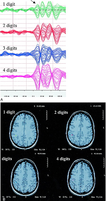
  • fig 2.
    • Download figure
    • Open in new tab
    • Download powerpoint
    fig 2.

    Magnetoencephalography waveforms and source localization.

    A, Representative magnetoencephalography waveforms for one, two, three, and four digits, for a single participant, single session. Waveforms are shown from the 37 sensor channels, collapsed onto a single horizontal time axis, ranging from 150 ms before stimulation to 150 ms after stimulation, capturing the initial evoked response. The y axis of these plots is a measure of evoked magnetic field amplitude, and all plots are shown to scale (50 fT/vertical division). All have recognizable peaks occurring approximately 30 to 40 ms after stimulus onset (arrow), with subsequent activity that is not the focus of this study. The amplitude of this peak seems to increase, somewhat monotonically, with an increasing number of stimulated digits.

    B, Source localization (single equivalent dipole modeling) of this magnetoencephalography-detected peak onto high resolution MR images (SPGR; 38/6/1; flip angle, 35°), to form magnetic source images (MSI) for one-, two-, three-, and four-digit stimulation shows the source estimate (colored point) to lie in the postcentral gyrus, which is expected to contain primary somatosensory representations. The ring surrounding the modeled current source indicates the 95% confidence limit for model/data fit. The 95% confidence volumes (from 3D ellipsoids of confidence) for the example in the figure are 0.17, 0.06, 0.08, and 0.02 cm3, respectively, for one-, two-, three-, and four-digit stimulation.

  • fig 3.
    • Download figure
    • Open in new tab
    • Download powerpoint
    fig 3.

    Variation of derived measures of the extent of cortical activation with increasing numbers of stimulated digits: intra- and interparticipant.

    A, Area of activation derived by functional MR imaging shows considerable intraparticipant variability and no trend toward increasing areas of activation with increasing numbers of stimulated digits, for averaged data for each individual. Measures of cortical activation are shown as a function of increasing numbers of stimulated digits with intraparticipant variability represented by the dispersion of like-colored dots. Mean individual participant trends are shown as solid lines.

    B, Evoked field amplitude as determined by magnetoencephalography. Despite overlap in the range of evoked fields elicited by each stimulation type across participants, the data from each individual are relatively clustered for each stimulation type; clear trends indicating increased activation with increased numbers of stimulated digits are seen for every individual participant. Measures of cortical activation are shown as a function of increasing numbers of stimulated digits with intraparticipant variability represented by the dispersion of like-colored dots. Mean individual participant trends are shown as solid lines.

    C, There is no evident correlation between functional MR imaging and magnetoencephalographic measures.

  • fig 4.
    • Download figure
    • Open in new tab
    • Download powerpoint
    fig 4.

    Interrater comparison for functional MR imaging. Despite differing sensitivities to activation identification (the slope of the correlation plot is approximately 0.6), the correlation between readers is strong (r ;eq 0.93), suggesting that observations and derived conclusions are not sensitive to arbitrary reader bias in definition of region of interest

Tables

  • Figures
  • Table1

    Statistical Significance of fMRI and MEG Measures of Cortical Extent of Activation

PreviousNext
Back to top

In this issue

American Journal of Neuroradiology
Vol. 21, Issue 8
1 Sep 2000
  • Table of Contents
  • Index by author
Advertisement
Print
Download PDF
Email Article

Thank you for your interest in spreading the word on American Journal of Neuroradiology.

NOTE: We only request your email address so that the person you are recommending the page to knows that you wanted them to see it, and that it is not junk mail. We do not capture any email address.

Enter multiple addresses on separate lines or separate them with commas.
Quantification and Reproducibility of Tracking Cortical Extent of Activation by Use of Functional MR Imaging and Magnetoencephalography
(Your Name) has sent you a message from American Journal of Neuroradiology
(Your Name) thought you would like to see the American Journal of Neuroradiology web site.
CAPTCHA
This question is for testing whether or not you are a human visitor and to prevent automated spam submissions.
Cite this article
Timothy P.L. Roberts, Elizabeth A. Disbrow, Heidi C. Roberts, Howard A. Rowley
Quantification and Reproducibility of Tracking Cortical Extent of Activation by Use of Functional MR Imaging and Magnetoencephalography
American Journal of Neuroradiology Sep 2000, 21 (8) 1377-1387;

Citation Manager Formats

  • BibTeX
  • Bookends
  • EasyBib
  • EndNote (tagged)
  • EndNote 8 (xml)
  • Medlars
  • Mendeley
  • Papers
  • RefWorks Tagged
  • Ref Manager
  • RIS
  • Zotero
0 Responses
Respond to this article
Share
Bookmark this article
Quantification and Reproducibility of Tracking Cortical Extent of Activation by Use of Functional MR Imaging and Magnetoencephalography
Timothy P.L. Roberts, Elizabeth A. Disbrow, Heidi C. Roberts, Howard A. Rowley
American Journal of Neuroradiology Sep 2000, 21 (8) 1377-1387;
del.icio.us logo Twitter logo Facebook logo Mendeley logo
  • Tweet Widget
  • Facebook Like
  • Google Plus One

Jump to section

  • Article
    • Abstract
    • Methods
    • Results
    • Discussion
    • Conclusion
    • Acknowledgments
    • Footnotes
    • References
  • Figures & Data
  • Info & Metrics
  • Responses
  • References
  • PDF

Related Articles

  • No related articles found.
  • PubMed
  • Google Scholar

Cited By...

  • Cortical reorganization in malformations of cortical development: A magnetoencephalographic study
  • Crossref
  • Google Scholar

This article has not yet been cited by articles in journals that are participating in Crossref Cited-by Linking.

More in this TOC Section

  • Enhanced Axonal Metabolism during Early Natalizumab Treatment in Relapsing-Remitting Multiple Sclerosis
  • Progression of Microstructural Damage in Spinocerebellar Ataxia Type 2: A Longitudinal DTI Study
  • SWI or T2*: Which MRI Sequence to Use in the Detection of Cerebral Microbleeds? The Karolinska Imaging Dementia Study
Show more BRAIN

Similar Articles

Advertisement

Indexed Content

  • Current Issue
  • Accepted Manuscripts
  • Article Preview
  • Past Issues
  • Editorials
  • Editor's Choice
  • Fellows' Journal Club
  • Letters to the Editor
  • Video Articles

Cases

  • Case Collection
  • Archive - Case of the Week
  • Archive - Case of the Month
  • Archive - Classic Case

More from AJNR

  • Trainee Corner
  • Imaging Protocols
  • MRI Safety Corner
  • Book Reviews

Multimedia

  • AJNR Podcasts
  • AJNR Scantastics

Resources

  • Turnaround Time
  • Submit a Manuscript
  • Submit a Video Article
  • Submit an eLetter to the Editor/Response
  • Manuscript Submission Guidelines
  • Statistical Tips
  • Fast Publishing of Accepted Manuscripts
  • Graphical Abstract Preparation
  • Imaging Protocol Submission
  • Evidence-Based Medicine Level Guide
  • Publishing Checklists
  • Author Policies
  • Become a Reviewer/Academy of Reviewers
  • News and Updates

About Us

  • About AJNR
  • Editorial Board
  • Editorial Board Alumni
  • Alerts
  • Permissions
  • Not an AJNR Subscriber? Join Now
  • Advertise with Us
  • Librarian Resources
  • Feedback
  • Terms and Conditions
  • AJNR Editorial Board Alumni

American Society of Neuroradiology

  • Not an ASNR Member? Join Now

© 2025 by the American Society of Neuroradiology All rights, including for text and data mining, AI training, and similar technologies, are reserved.
Print ISSN: 0195-6108 Online ISSN: 1936-959X

Powered by HighWire