
Article Figures & Data
Figures
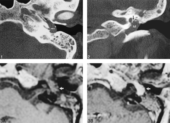
- fig 1.
Axial unenhanced CT scan shows a well-circumscribed mass (M) in the tympanic cavity. Note the erosion of the cochlear promontory (arrow).fig 2. Coronal unenhanced CT scan shows the mass confined to the tympanic cavity. Note the normal-sized tympanic segment of the facial nerve canal (arrow).fig 3. A, Axial unenhanced T1-weighted image shows a tympanic mass (arrow) that is isointense to brain parenchyma (500/10/2 [TR/TE/excitations]); B, Axial contrast-enhanced T1-weighted image reveals intense, homogeneous enhancement of the mass (arrow) (500/10/2)
- fig 4.
Axial photomicrograph of a normal temporal bone specimen shows the Jacobson's nerve adjacent to the cochlear promontory (arrow). Note the stapes footplate (open arrow), and the facial nerve (F).fig 5. Axial unenhanced CT scan shows normal-sized inferior canaliculus (arrows). Note the glomus typanicum (arrowhead)


In this issue


Jump to section
Related Articles
Cited By...
- No citing articles found.