
Article Figures & Data
Figures
- fig 1.
A, Cross section through the mesencephalon at the level of the red nucleus shows the subdivision of the parvicellular subnucleus into the pars oralis and pars dorsomedialis (Heidenhain stain; original magnification × 8).
B, Cross section through the mesencephalon at the level of the red nucleus shows the subdivision of the parvicellular subnucleus into the pars oralis and pars dorsomedialis (original magnification × 30). The corresponding area of the adjacent myelin-stained section is delineated by the square in A.
C, Drawing of red nucleus partitions according to A shows the medullary lamella (ML) in the middle of the parvicellular subnucleus of the red nucleus.
Aq indicates sylvian aqueduct; Dm, pars dorsomedialis; Or, pars oralis; Mm, mammillary body; Su.n, substantia nigra. Parts A and B were reproduced with permission from S. Karger, Basel, Switzerland.
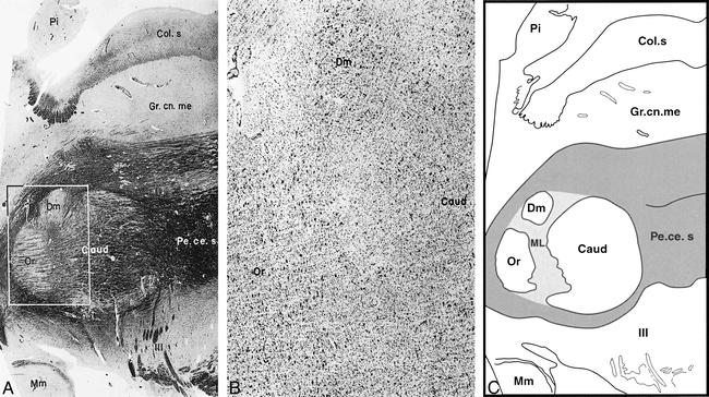
- fig 2.
A, Sagittal section through the mesencephalon at the level of the red nucleus shows the subdivision of the parvicellular subnucleus into the pars oralis, pars dorsomedialis, and pars caudalis (Heidenhain stain; original magnification × 8).
B, Sagittal section through the mesencephalon at the level of the red nucleus shows the subdivision of the parvicellular subnucleus into the pars oralis, pars dorsomedialis, and pars caudalis (original magnification × 30). The corresponding area of the adjacent myelin-stained section is delineated by the square in A.
C, Drawing of red nucleus partitions according to A shows the medullary lamellae (ML) in the middle of the parvicellular subnucleus of the red nucleus.
Dm indicates pars dorsomedialis; Or, pars oralis; Caud, pars caudalis; Mm, mammillary body; Pe.ce.s, pedunculus cerebellaris superior; Pi, pineal body; III, rootlet of the oculomotor nerve; Col.s, colliculus superior; Gr. cn.me, griseum centrale mesencephli. Parts A and B were reproduced with permission from S. Karger, Basel, Switzerland.
- fig 3.
A, Typical oblique axial gradient-echo MR image (60/40/15) for one subject through the centers of the red and dentate nuclei of both sides. A lamella (arrow) within the red nuclei of both sides is clearly shown. It is triangular in shape with the apex directed medially. The signal intensity of the lamella is relatively higher than that of the other parts of the red nucleus. B, Enlarged view of the red nuclei. Arrow indicates the medullary lamella
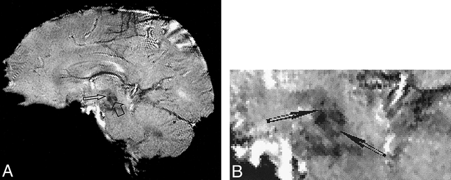
- fig 4.
A, Typical oblique sagittal gradient-echo MR image (60/40/15) for one subject through the centers of the right red and dentate nuclei. Two lamellae (arrows) within the right red nucleus are clearly shown as cross-shaped structures. The signal intensity of the lamellae is relatively higher than that of the other parts of the red nucleus.
B, Enlarged view of the red nuclei. Arrows indicate the medullary lamellae.
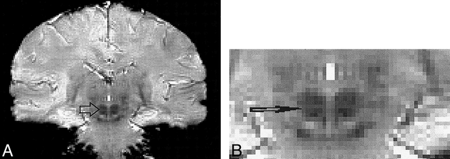
- fig 5.
A, Typical coronal gradient-echo MR image (60/40/15) for one subject through both red nuclei. A lamella (arrow) is clearly seen within the nuclei extending lateromedially throughout the red nucleus. The signal intensity of the lamella is relatively higher than that of the other parts of the red nucleus.
B, Enlarged view of the red nuclei. Arrow indicates the medullary lamella.


