
Article Figures & Data
Figures
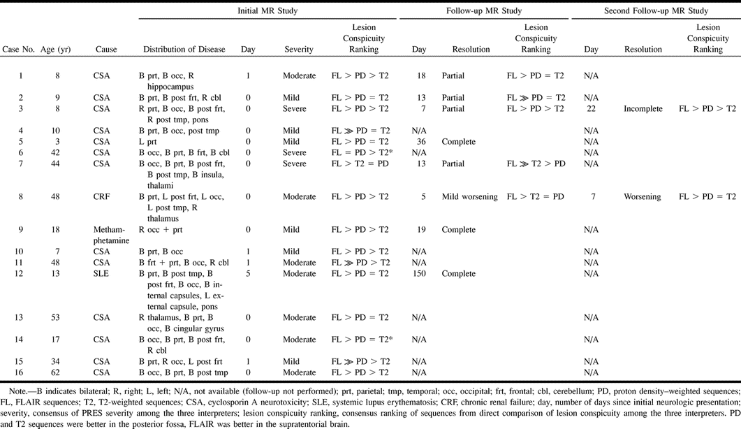
- fig 1.
Case 12: 13-year-old girl with systemic lupus erythematosus who presented with hypertension and status epilepticus. MR imaging was performed 5 days later.
A–C, FLAIR axial sections show extensive subcortical white matter edema bilaterally (arrows) with only minimal cortical involvement. Lesions are in the parietal, occipital, posterior frontal, and posterior temporal lobes and in the left corona radiata (asterisk). Additional T2 hyperintensity is seen in the white matter around the lentiform nuclei (arrowheads). Findings are consistent with typical changes of PRES, especially in the parietooccipital regions, and were considered moderate (severity index = 2) for the purposes of this study.
D and E, Follow-up MR study obtained 150 days after initial neurologic examination reveals complete resolution of lesions on FLAIR images.
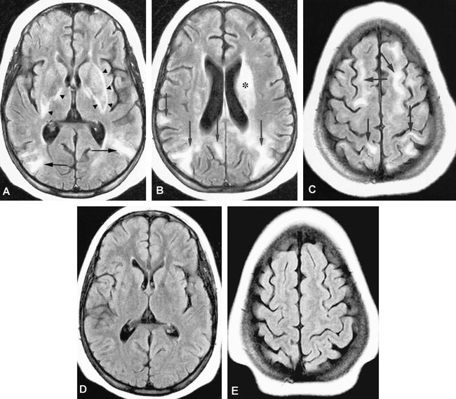
- fig 2.
Case 8: 48-year-old woman with history of vasculitis and chronic renal failure who presented with seizures.
A, Initial MR study, FLAIR sequence, reveals typical subcortical edema of PRES in the left posterior frontal lobe (arrow). This patient also had mild cortical involvement of the parietooccipital regions, the left posterior temporal lobe, and the left thalamus. These findings were assigned a severity ranking of 2 (moderate disease).
B, Follow-up FLAIR image 5 days after original presentation. Although the left posterior frontal subcortical edema has resolved, there was an overall progression of the findings of PRES with new right posterior frontal cortical hyperintensity (arrows).
C, FLAIR image, at same level, 7 days after original presentation, shows mild worsening of edema, now bifrontal, with some new subcortical foci (arrowheads).
D, T2-weighted image shows new bilateral cerebellar hyperintense foci (arrows).
E, Cerebellar lesions are seen better on FLAIR image (arrows). This patient has parietooccipital lesions typical of severe PRES. Extension of edema to involve the cerebellum bilaterally was considered to warrant a severity index of 3 (severe disease).
- fig 3.
Case 4: 10-year-old boy being treated with CSA for bone marrow transplantation for acute lymphocytic leukemia. Shortly before imaging, the patient had two episodes of focal seizures and acute hypertension coincident with seizure activity. The serum CSA level was normal at the time the seizures occurred. Before the day of imaging, the patient had been normotensive.
A, Axial proton density–weighted image.
B, Axial T2-weighted image at same level.
C, FLAIR image at same level as A and B reveals cortical T2 hyperintensity in a gyral pattern bilaterally in the occipitoparietal lobes (arrows).
D, Axial FLAIR image, superior to A, suggests additional subtle cortical hyperintensity along gyri of the left parietal and posterior frontal lobes (arrows). This type of subtle cortical involvement was given a severity rating of 1 (mild disease).
- fig 4.
Case 11: 48-year-old man being treated with CSA who presented with generalized seizures shortly before imaging.
A and B, Proton density–weighted (A) and axial T2-weighted (B) sections reveal a nonspecific punctate white matter hyperintensity (arrow) in the left parietal lobe.
C, Corresponding turbo-FLAIR image shows biparietal increased signal within the cortex (white arrows) and subcortical white matter (black arrow), representing edema and suggesting the radiologic diagnosis of PRES.
D and E, Adjacent proton density–weighted (D) and T2-weighted (E) images, one level superiorly, show increased cortical (white arrows) and subcortical white matter signal due to edema.
F, Turbo-FLAIR image at same level again reveals the cortical-based hyperintensities that are difficult to appreciate on standard dual-echo sequences, owing to the adjacent bright CSF. FLAIR unmasks this abnormality by suppressing CSF signal. This case was assigned a moderate severity score on the basis of the FLAIR imaging findings, although on the basis of proton density–or T2-weighted images it would have been considered mild.
- fig 5.
Mean of the percentage of PRES lesion distribution with respect to gray and white matter. Mean percentages were also calculated for each severity subgroup and for all cases. There was a trend toward greater white matter involvement with increasing disease severity. The overall mean percentage of lesions was 46% in the gray matter and 54% in the white matter


Tables

Comparison of MR sequences in 16 patients with radiologic and clinical findings of posterior reversible encephalopathy syndrome
In this issue


Jump to section
Related Articles
Cited By...
- Posterior reversible encephalopathy syndrome (PRES): diagnosis and management
- Pediatric Acute Toxic Leukoencephalopathy: Prediction of the Clinical Outcome by FLAIR and DWI for Various Etiologies
- Posterior reversible encephalopathy syndrome associated with the use of chemotherapeutic agents: a rare complication after treatment with vinorelbine
- Hypertensive brainstem encephalopathy: a diagnosis often overlooked
- Posterior reversible encephalopathy syndrome due to hypercalcaemia: a rare cause
- Acute Toxic Leukoencephalopathy: Etiologies, Imaging Findings, and Outcomes in 101 Patients
- Controversy of posterior reversible encephalopathy syndrome: what have we learnt in the last 20 years?
- Reply:
- Utility and Significance of Gadolinium-Based Contrast Enhancement in Posterior Reversible Encephalopathy Syndrome
- Neuroimaging Features and Predictors of Outcome in Eclamptic Encephalopathy: A Prospective Observational Study
- Reversible Posterior Leukoencephalopathy Syndrome and Bevacizumab in Breast Cancer
- Detection of Microhemorrhage in Posterior Reversible Encephalopathy Syndrome Using Susceptibility-Weighted Imaging
- The posterior reversible encephalopathy syndrome: what's certain, what's new?
- Posterior reversible encephalopathy syndrome: long-term follow-up
- A phase 2 multiple ascending dose trial of bapineuzumab in mild to moderate Alzheimer disease
- POSTERIOR REVERSIBLE ENCEPHALOPATHY SYNDROME IN NEUROMYELITIS OPTICA SPECTRUM DISORDERS
- Headache, blindness and a seizure after childbirth
- Posterior reversible encephalopathy syndrome in SLE nephritis
- Gemcitabine-Induced Reversible Posterior Leukoencephalopathy Syndrome: A Case Report and Review of the Literature
- Seizures following bone marrow transplantation
- Reversible diffusion MRI abnormalities and transient mutism after liver transplantation
- Reversible diffusion MRI abnormalities and transient mutism after liver transplantation
- Exclusion of brain lesions: is MR contrast medium required after a negative fluid-attenuated inversion recovery sequence?
- Nitroglycerin-aggravated pre-eclamptic posterior reversible encephalopathy syndrome (PRES)
- Acute headache as a presenting symptom of tacrolimus encephalopathy
- Don't throw in the towel! A case of reversible coma
- ""T2 Washout"": An Explanation for Normal Diffusion-Weighted Images Despite Abnormal Apparent Diffusion Coefficient Maps
- Quantitative Assessment of Diffusion Abnormalities in Posterior Reversible Encephalopathy Syndrome