Advertisement

Main menu

  • Home
  • Content
    • Current Issue
    • Accepted Manuscripts
    • Article Preview
    • Past Issue Archive
    • Video Articles
    • AJNR Case Collection
    • Case of the Week Archive
    • Case of the Month Archive
    • Classic Case Archive
  • Special Collections
    • AJNR Awards
    • Low-Field MRI
    • Alzheimer Disease
    • ASNR Foundation Special Collection
    • Photon-Counting CT
    • View All
  • Multimedia
    • AJNR Podcasts
    • AJNR SCANtastic
    • Trainee Corner
    • MRI Safety Corner
    • Imaging Protocols
  • For Authors
    • Submit a Manuscript
    • Submit a Video Article
    • Submit an eLetter to the Editor/Response
    • Manuscript Submission Guidelines
    • Statistical Tips
    • Fast Publishing of Accepted Manuscripts
    • Graphical Abstract Preparation
    • Imaging Protocol Submission
    • Author Policies
  • About Us
    • About AJNR
    • Editorial Board
    • Editorial Board Alumni
  • More
    • Become a Reviewer/Academy of Reviewers
    • Subscribers
    • Permissions
    • Alerts
    • Feedback
    • Advertisers
    • ASNR Home

User menu

  • Alerts
  • Log in

Search

  • Advanced search
American Journal of Neuroradiology

American Journal of Neuroradiology

ASHNR American Society of Functional Neuroradiology ASHNR American Society of Pediatric Neuroradiology ASSR
  • Alerts
  • Log in

Advanced Search

  • Home
  • Content
    • Current Issue
    • Accepted Manuscripts
    • Article Preview
    • Past Issue Archive
    • Video Articles
    • AJNR Case Collection
    • Case of the Week Archive
    • Case of the Month Archive
    • Classic Case Archive
  • Special Collections
    • AJNR Awards
    • Low-Field MRI
    • Alzheimer Disease
    • ASNR Foundation Special Collection
    • Photon-Counting CT
    • View All
  • Multimedia
    • AJNR Podcasts
    • AJNR SCANtastic
    • Trainee Corner
    • MRI Safety Corner
    • Imaging Protocols
  • For Authors
    • Submit a Manuscript
    • Submit a Video Article
    • Submit an eLetter to the Editor/Response
    • Manuscript Submission Guidelines
    • Statistical Tips
    • Fast Publishing of Accepted Manuscripts
    • Graphical Abstract Preparation
    • Imaging Protocol Submission
    • Author Policies
  • About Us
    • About AJNR
    • Editorial Board
    • Editorial Board Alumni
  • More
    • Become a Reviewer/Academy of Reviewers
    • Subscribers
    • Permissions
    • Alerts
    • Feedback
    • Advertisers
    • ASNR Home
  • Follow AJNR on Twitter
  • Visit AJNR on Facebook
  • Follow AJNR on Instagram
  • Join AJNR on LinkedIn
  • RSS Feeds

AJNR Awards, New Junior Editors, and more. Read the latest AJNR updates

Research ArticleSpine Imaging and Spine Image-Guided Interventions

Partial Aplasia of the Posterior Arch of the Atlas with an Isolated Posterior Arch Remnant: Findings in Three Cases

Aseem Sharma, Shailesh B. Gaikwad, Parminder S. Deol, Nalin K. Mishra and Shashank S. Kale
American Journal of Neuroradiology June 2000, 21 (6) 1167-1171;
Aseem Sharma
  • Find this author on Google Scholar
  • Find this author on PubMed
  • Search for this author on this site
Shailesh B. Gaikwad
  • Find this author on Google Scholar
  • Find this author on PubMed
  • Search for this author on this site
Parminder S. Deol
  • Find this author on Google Scholar
  • Find this author on PubMed
  • Search for this author on this site
Nalin K. Mishra
  • Find this author on Google Scholar
  • Find this author on PubMed
  • Search for this author on this site
Shashank S. Kale
  • Find this author on Google Scholar
  • Find this author on PubMed
  • Search for this author on this site
  • Article
  • Figures & Data
  • Info & Metrics
  • Responses
  • References
  • PDF
Loading

Article Figures & Data

Figures

  • Tables
  • fig 1.
    • Download figure
    • Open in new tab
    • Download powerpoint
    fig 1.

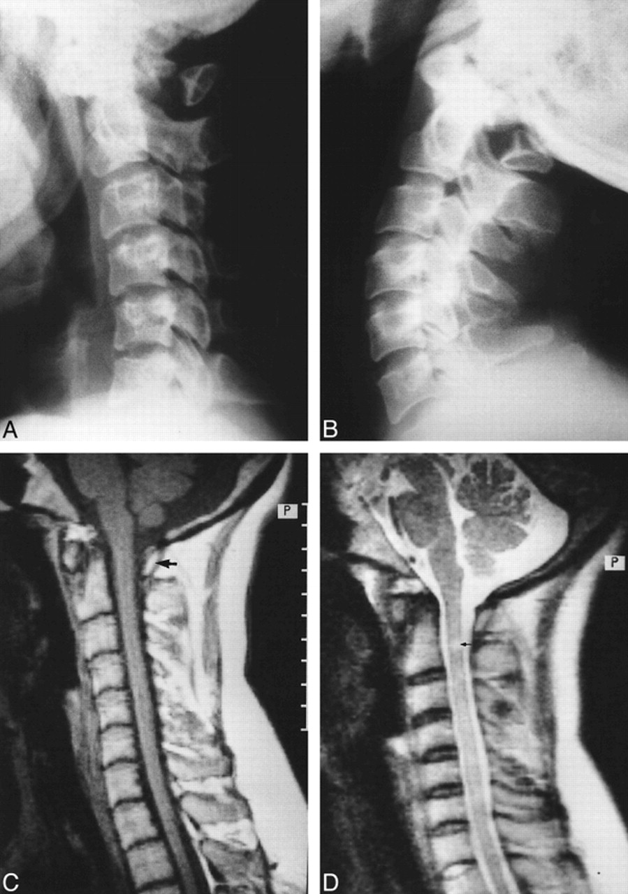
    32-year-old woman with intermittent weakness and numbness of all four limbs.

    A and B, Lateral radiographs of cervical spine taken in flexion (A) and extension (B) reveal partial aplasia of the posterior arch of the atlas with an isolated posterior bony fragment. Note the anterior displacement of the bony fragment during extension (B).

    C, Midsagittal T1-weighted MR image through the cervical spine shows no compression of the cord by the posterior bony fragment (arrow).

    D, Corresponding T2-weighted image shows a small, localized region of hyperintensity within the cord (arrow). Note that the signal alteration is seen slightly inferior to the anteroinferior margin of the isolated posterior arch remnant. A superimposed canal stenosis was noted from C2 through C5.

  • fig 2.
    • Download figure
    • Open in new tab
    • Download powerpoint
    fig 2.

    35-year-old woman with neck pain and paresthesias in both upper limbs.

    A and B, In varying degrees of extension, plain radiographs show partial aplasia of the posterior arch of the atlas, with an isolated posterior tubercle. No obvious anterior displacement of the posterior tubercle is observed during extension.

    C, Midsagittal T1-weighted MR image reveals no cord compression.

    D, Corresponding T2-weighted image shows small, focal intramedullary hyperintensity (arrow) located just below the level of the posterior tubercle. In addition, there is focal canal stenosis at the C2–C3 disk level.

  • fig 3.
    • Download figure
    • Open in new tab
    • Download powerpoint
    fig 3.

    30-year-old man with neck pain and occasional upper limb paresthesias.

    A and B, Flexion (A) and extension (B) plain radiographs of the cervical spine show partial aplasia of the posterior arch of the atlas with an isolated posterior arch remnant. The isolated posterior fragment moves anteriorly into the spinal canal during extension (B).

    C and D, Midsagittal T1-weighted (C) and T2-weighted (D) MR images reveal no compression or signal alteration of the spinal cord. Significant canal stenosis from C3 to C7 is present along with a superimposed disk bulge/protrusion at C5–C6.

Tables

  • Figures
  • Table1

    Classification of congenital anomalies of the posterior arch of the atlas according to Curriano (14)

PreviousNext
Back to top

In this issue

American Journal of Neuroradiology
Vol. 21, Issue 6
1 Jun 2000
  • Table of Contents
  • Index by author
Advertisement
Print
Download PDF
Email Article

Thank you for your interest in spreading the word on American Journal of Neuroradiology.

NOTE: We only request your email address so that the person you are recommending the page to knows that you wanted them to see it, and that it is not junk mail. We do not capture any email address.

Enter multiple addresses on separate lines or separate them with commas.
Partial Aplasia of the Posterior Arch of the Atlas with an Isolated Posterior Arch Remnant: Findings in Three Cases
(Your Name) has sent you a message from American Journal of Neuroradiology
(Your Name) thought you would like to see the American Journal of Neuroradiology web site.
CAPTCHA
This question is for testing whether or not you are a human visitor and to prevent automated spam submissions.
Cite this article
Aseem Sharma, Shailesh B. Gaikwad, Parminder S. Deol, Nalin K. Mishra, Shashank S. Kale
Partial Aplasia of the Posterior Arch of the Atlas with an Isolated Posterior Arch Remnant: Findings in Three Cases
American Journal of Neuroradiology Jun 2000, 21 (6) 1167-1171;

Citation Manager Formats

  • BibTeX
  • Bookends
  • EasyBib
  • EndNote (tagged)
  • EndNote 8 (xml)
  • Medlars
  • Mendeley
  • Papers
  • RefWorks Tagged
  • Ref Manager
  • RIS
  • Zotero
0 Responses
Respond to this article
Share
Bookmark this article
Partial Aplasia of the Posterior Arch of the Atlas with an Isolated Posterior Arch Remnant: Findings in Three Cases
Aseem Sharma, Shailesh B. Gaikwad, Parminder S. Deol, Nalin K. Mishra, Shashank S. Kale
American Journal of Neuroradiology Jun 2000, 21 (6) 1167-1171;
del.icio.us logo Twitter logo Facebook logo Mendeley logo
  • Tweet Widget
  • Facebook Like
  • Google Plus One

Jump to section

  • Article
    • Abstract
    • Case Reports
    • Discussion
    • Conclusion
    • Footnotes
    • References
  • Figures & Data
  • Info & Metrics
  • Responses
  • References
  • PDF

Related Articles

  • No related articles found.
  • PubMed
  • Google Scholar

Cited By...

  • Republished: Congenital anomaly of the posterior arch of the atlas: a rare risk factor for posterior circulation stroke
  • Delayed diagnosis of fractured anterior arch of the atlas in a young child
  • Congenital anomaly of the posterior arch of the atlas: a rare risk factor for posterior circulation stroke
  • Congenital absence of the posterior arch of the atlas associated with a fracture of the anterior arch
  • Cervical radiography
  • Crossref
  • Google Scholar

This article has not yet been cited by articles in journals that are participating in Crossref Cited-by Linking.

More in this TOC Section

  • Cone Beam CT Myelography
  • Cone Beam CTM for detecting CSF-venous Fistulas
  • Post-Procedural Brachial Neuritis Features
Show more Spine Imaging and Spine Image-Guided Interventions

Similar Articles

Advertisement

Indexed Content

  • Current Issue
  • Accepted Manuscripts
  • Article Preview
  • Past Issues
  • Editorials
  • Editor's Choice
  • Fellows' Journal Club
  • Letters to the Editor
  • Video Articles

Cases

  • Case Collection
  • Archive - Case of the Week
  • Archive - Case of the Month
  • Archive - Classic Case

More from AJNR

  • Trainee Corner
  • Imaging Protocols
  • MRI Safety Corner

Multimedia

  • AJNR Podcasts
  • AJNR Scantastics

Resources

  • Turnaround Time
  • Submit a Manuscript
  • Submit a Video Article
  • Submit an eLetter to the Editor/Response
  • Manuscript Submission Guidelines
  • Statistical Tips
  • Fast Publishing of Accepted Manuscripts
  • Graphical Abstract Preparation
  • Imaging Protocol Submission
  • Evidence-Based Medicine Level Guide
  • Publishing Checklists
  • Author Policies
  • Become a Reviewer/Academy of Reviewers
  • News and Updates

About Us

  • About AJNR
  • Editorial Board
  • Editorial Board Alumni
  • Alerts
  • Permissions
  • Not an AJNR Subscriber? Join Now
  • Advertise with Us
  • Librarian Resources
  • Feedback
  • Terms and Conditions
  • AJNR Editorial Board Alumni

American Society of Neuroradiology

  • Not an ASNR Member? Join Now

© 2025 by the American Society of Neuroradiology All rights, including for text and data mining, AI training, and similar technologies, are reserved.
Print ISSN: 0195-6108 Online ISSN: 1936-959X

Powered by HighWire