
Article Figures & Data
Figures
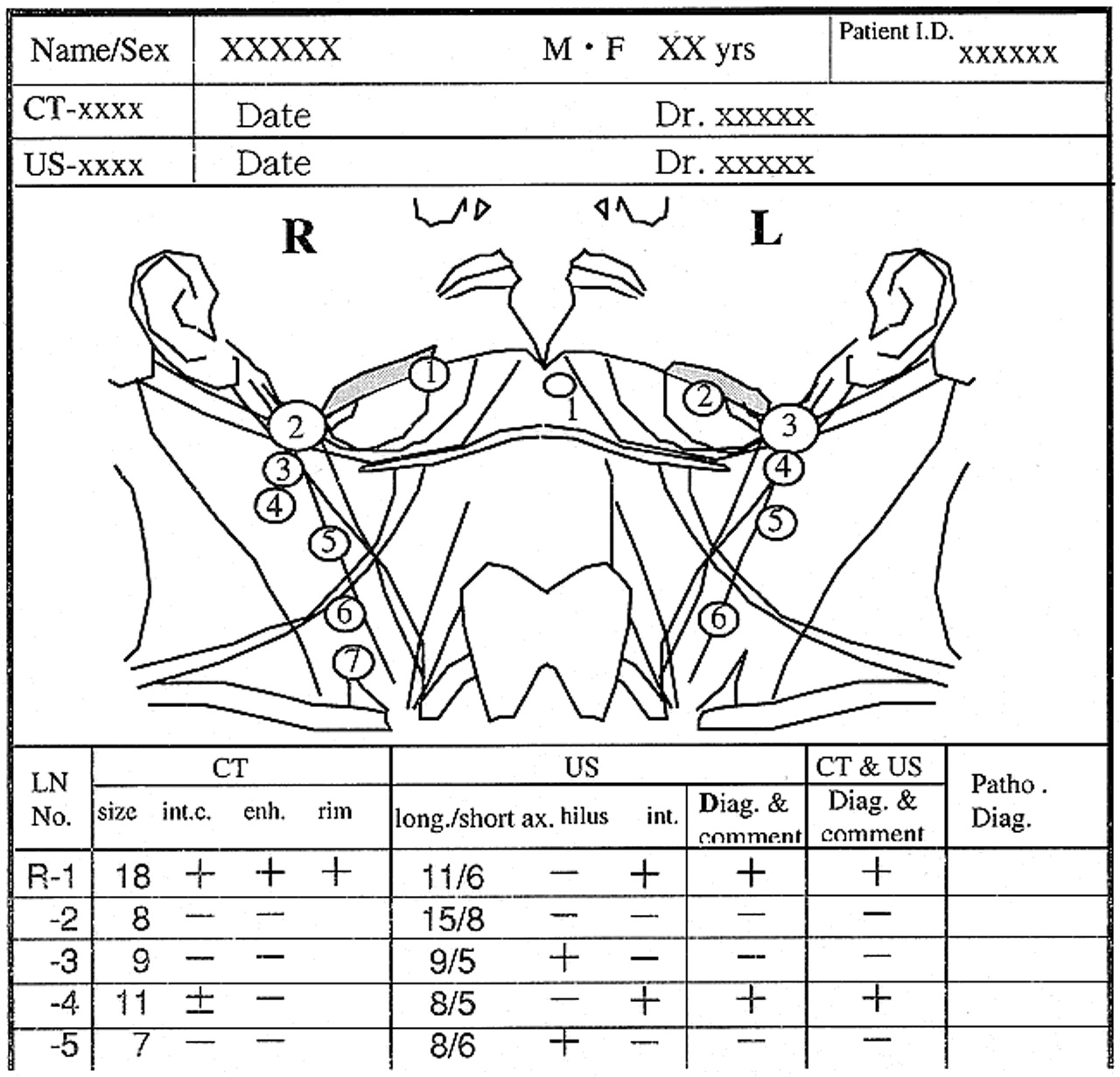
- fig 1.
An example of a report of a cervical lymph node with topographic illustrations
- fig 2.
Follow-up sonogram of lymph node in patient 2. A, The first follow-up sonogram. The lymph node measures 3 mm in short-axis diameter. B, The second follow-up sonogram (28 days after the first follow-up sonogram was obtained). Short-axis diameter of the lymph node has increased by 2 mm. Slight, internal echogenicity, suspicious for metastasis, is visible. C, The third follow-up sonogram (9 days after the second follow-up sonogram was obtained). Short-axis diameter of the lymph node has increased by 1 mm. Marked internal echogenicity is increased, and the boundary of the node is unclear, suggesting extranodal extension of disease. Pathologic examination of this lymph node confirmed metastasis, with extranodal extension of disease.
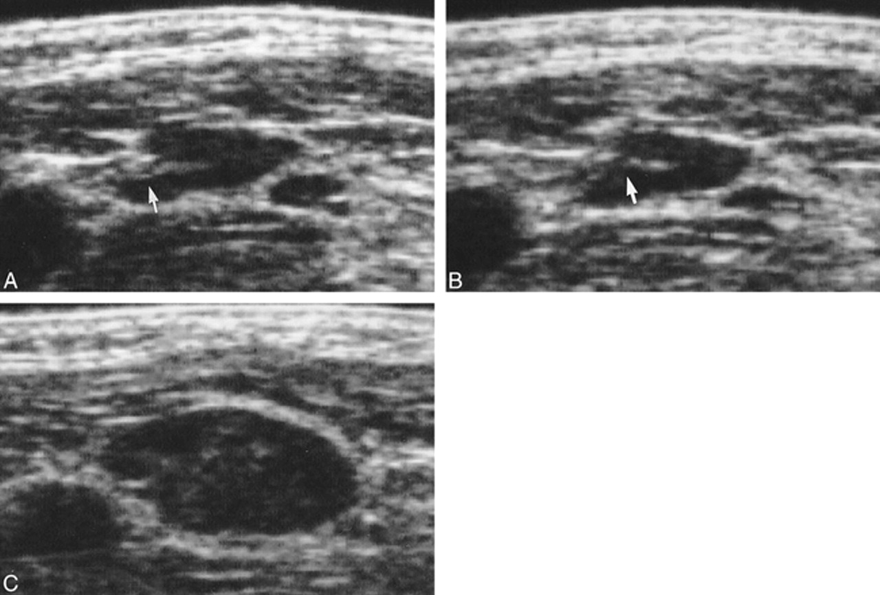
- fig 3.
Follow-up sonograms of the lymph node in patient 5. A, The third follow-up sonogram. The lymph node measures 5 mm in short-axis diameter, with normal hilar echoes (arrow). B, The fourth follow-up sonogram (35 days after the third follow-up sonogram was obtained). The size of the lymph node has not changed, and normal hilar echoes are also observed. In retrospect, we should have noticed that the hilar echoes were slightly less distinct compared with those revealed by the last sonogram (arrow), and should have performed sonography at a shorter interval after performing the third follow-up examination. C, The fifth follow-up sonogram (42 days after the fourth follow-up sonogram was obtained). The lymph node has increased by 5 mm in short-axis diameter. Internal echogenicity is also observed, and the normal hilar echoes are no longer visible. Pathologic anlaysis of this lymph node confirmed metastasis with extranodal extension of disease.


Tables

The changes of short-axis diameter and internal structures of metastatic lymph nodes in follow-up sonography


