Advertisement

Main menu

  • Home
  • Content
    • Current Issue
    • Accepted Manuscripts
    • Article Preview
    • Past Issue Archive
    • Video Articles
    • AJNR Case Collection
    • Case of the Week Archive
    • Case of the Month Archive
    • Classic Case Archive
  • Special Collections
    • AJNR Awards
    • Low-Field MRI
    • Alzheimer Disease
    • ASNR Foundation Special Collection
    • Photon-Counting CT
    • View All
  • Multimedia
    • AJNR Podcasts
    • AJNR SCANtastic
    • Trainee Corner
    • MRI Safety Corner
    • Imaging Protocols
  • For Authors
    • Submit a Manuscript
    • Submit a Video Article
    • Submit an eLetter to the Editor/Response
    • Manuscript Submission Guidelines
    • Statistical Tips
    • Fast Publishing of Accepted Manuscripts
    • Graphical Abstract Preparation
    • Imaging Protocol Submission
    • Author Policies
  • About Us
    • About AJNR
    • Editorial Board
    • Editorial Board Alumni
  • More
    • Become a Reviewer/Academy of Reviewers
    • Subscribers
    • Permissions
    • Alerts
    • Feedback
    • Advertisers
    • ASNR Home

User menu

  • Alerts
  • Log in

Search

  • Advanced search
American Journal of Neuroradiology

American Journal of Neuroradiology

ASHNR American Society of Functional Neuroradiology ASHNR American Society of Pediatric Neuroradiology ASSR
  • Alerts
  • Log in

Advanced Search

  • Home
  • Content
    • Current Issue
    • Accepted Manuscripts
    • Article Preview
    • Past Issue Archive
    • Video Articles
    • AJNR Case Collection
    • Case of the Week Archive
    • Case of the Month Archive
    • Classic Case Archive
  • Special Collections
    • AJNR Awards
    • Low-Field MRI
    • Alzheimer Disease
    • ASNR Foundation Special Collection
    • Photon-Counting CT
    • View All
  • Multimedia
    • AJNR Podcasts
    • AJNR SCANtastic
    • Trainee Corner
    • MRI Safety Corner
    • Imaging Protocols
  • For Authors
    • Submit a Manuscript
    • Submit a Video Article
    • Submit an eLetter to the Editor/Response
    • Manuscript Submission Guidelines
    • Statistical Tips
    • Fast Publishing of Accepted Manuscripts
    • Graphical Abstract Preparation
    • Imaging Protocol Submission
    • Author Policies
  • About Us
    • About AJNR
    • Editorial Board
    • Editorial Board Alumni
  • More
    • Become a Reviewer/Academy of Reviewers
    • Subscribers
    • Permissions
    • Alerts
    • Feedback
    • Advertisers
    • ASNR Home
  • Follow AJNR on Twitter
  • Visit AJNR on Facebook
  • Follow AJNR on Instagram
  • Join AJNR on LinkedIn
  • RSS Feeds

AJNR Awards, New Junior Editors, and more. Read the latest AJNR updates

Research ArticleBRAIN

The Influence of Gliomas and Nonglial Space-occupying Lesions on Blood-oxygen-level–dependent Contrast Enhancement

Axel Schreiber, Ulrich Hubbe, Sargon Ziyeh and Jürgen Hennig
American Journal of Neuroradiology June 2000, 21 (6) 1055-1063;
Axel Schreiber
  • Find this author on Google Scholar
  • Find this author on PubMed
  • Search for this author on this site
Ulrich Hubbe
  • Find this author on Google Scholar
  • Find this author on PubMed
  • Search for this author on this site
Sargon Ziyeh
  • Find this author on Google Scholar
  • Find this author on PubMed
  • Search for this author on this site
Jürgen Hennig
  • Find this author on Google Scholar
  • Find this author on PubMed
  • Search for this author on this site
  • Article
  • Figures & Data
  • Info & Metrics
  • Responses
  • References
  • PDF
Loading

Article Figures & Data

Figures

  • Tables
  • fig 1.
    • Download figure
    • Open in new tab
    • Download powerpoint
    fig 1.

    Adjacent to nonglial tumors, a normal volume of BOLD contrast enhancement was elicited.

    A, Case 4. Arrows point to a small chronic abscess in the left precentral gyrus within the hand area. Upper row (T2-weighted turbo spin-echo sequence: 4500/120/1) shows edema apparently confined to the white matter. A narrow band of cortex is spared (open arrowheads). Lower row (echo-planar image: 3500/84/1), shows BOLD contrast enhancement in the cortex directly adjacent to the edema (arrowheads). Neither the edema nor the lesion affect the BOLD contrast enhancement.

    B, Case 10. Arrows point to a metastatic lesion located in the right postcentral gyrus, slightly superior to the hand area. Arrowheads point to the functional activation of the sensorimotor cortex. Upper row is an overlay of the activation map onto the full head volume (MPRAGE: 9, 7/4/1 [TR/TE/excitations]). Lower row is an overlay onto the functional echo-planar sections (3500/84/1). In the ipsilateral hemisphere, the activation is squeezed between the swollen pre- and postcentral gyrus, but is not reduced. There is very little activation in the contralateral hemisphere.

  • fig 2.
    • Download figure
    • Open in new tab
    • Download powerpoint
    fig 2.

    Case 20. The patient was suffering from an anaplastic astrocytoma invading the precentral gyrus. She presented with focal epileptic seizures of the face on the left side but did not have a motor deficit. Overlay of the activation map onto the functional echo-planar image (3500/84/1) shows activation nearly symmetrical in the upper two sections lining the Ω-shaped hand-motor area. There is less activation in the ipsilateral sensorimotor cortex on the lower two sections. The signal hyperintense parts of the sensorimotor cortex do not display BOLD contrast enhancement. Region of interest 1 is the cluster of activated voxels in the most medial part of the pre- and postcentral gyrus. Region of interest 2 is on the lateral continuation of the pre- and postcentral gyrus within an area of T2 signal hyperintensity of the cerebral cortex. The signal hyperintensity likely indicates gliomatous infiltration; in this case, histologic analysis of the resected specimen showed diffuse infiltration of the cortex. The interval of the signal intensity of region of interest 1 shows task-related signal changes (BOLD contrast enhancement) of approximately 6%, with hemodynamic delays of one to three images (gray bars indicate stimulation measurements). Region of interest 2 does not show BOLD contrast enhancement, although the analogous area in the left hemisphere is strongly activated.

  • fig 3.
    • Download figure
    • Open in new tab
    • Download powerpoint
    fig 3.

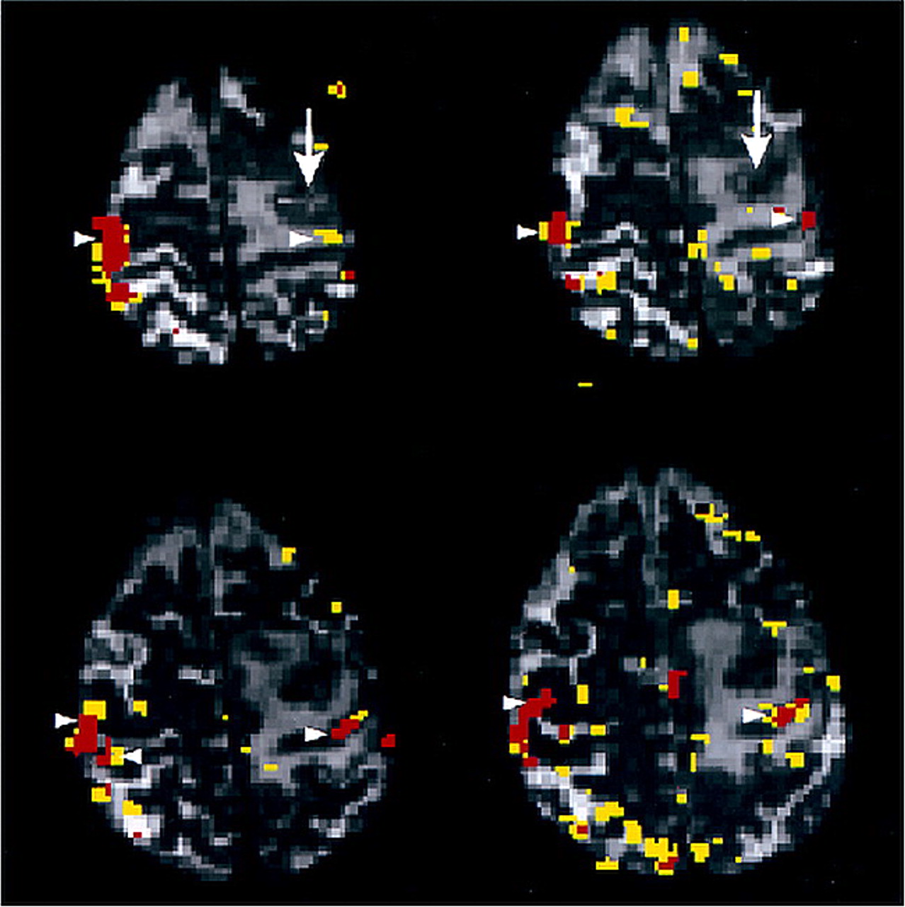
    Case 6. Arrows point to a metastasis of a neuroendocrine active lung cancer located in the left precentral gyrus near the hand area. A large edema extends into the pre- and postcentral gyrus. The patient presented with focal epileptic seizures starting at the right hand, generalizing secondarily. Arrowheads point to clusters of activation in the sensorimotor cortex overlaid onto the functional echo-planar sections (3500/84/1). On the ipsilateral hemisphere, the functional activation is far less than on the contralateral hemisphere. The parieto-occipital clusters on the lowest section are attributable to head motion

Tables

  • Figures
  • Table1

    Results and characterization of space-occupying lesions in patients who underwent successful FMR imaging

PreviousNext
Back to top

In this issue

American Journal of Neuroradiology
Vol. 21, Issue 6
1 Jun 2000
  • Table of Contents
  • Index by author
Advertisement
Print
Download PDF
Email Article

Thank you for your interest in spreading the word on American Journal of Neuroradiology.

NOTE: We only request your email address so that the person you are recommending the page to knows that you wanted them to see it, and that it is not junk mail. We do not capture any email address.

Enter multiple addresses on separate lines or separate them with commas.
The Influence of Gliomas and Nonglial Space-occupying Lesions on Blood-oxygen-level–dependent Contrast Enhancement
(Your Name) has sent you a message from American Journal of Neuroradiology
(Your Name) thought you would like to see the American Journal of Neuroradiology web site.
CAPTCHA
This question is for testing whether or not you are a human visitor and to prevent automated spam submissions.
Cite this article
Axel Schreiber, Ulrich Hubbe, Sargon Ziyeh, Jürgen Hennig
The Influence of Gliomas and Nonglial Space-occupying Lesions on Blood-oxygen-level–dependent Contrast Enhancement
American Journal of Neuroradiology Jun 2000, 21 (6) 1055-1063;

Citation Manager Formats

  • BibTeX
  • Bookends
  • EasyBib
  • EndNote (tagged)
  • EndNote 8 (xml)
  • Medlars
  • Mendeley
  • Papers
  • RefWorks Tagged
  • Ref Manager
  • RIS
  • Zotero
0 Responses
Respond to this article
Share
Bookmark this article
The Influence of Gliomas and Nonglial Space-occupying Lesions on Blood-oxygen-level–dependent Contrast Enhancement
Axel Schreiber, Ulrich Hubbe, Sargon Ziyeh, Jürgen Hennig
American Journal of Neuroradiology Jun 2000, 21 (6) 1055-1063;
del.icio.us logo Twitter logo Facebook logo Mendeley logo
  • Tweet Widget
  • Facebook Like
  • Google Plus One

Jump to section

  • Article
    • Abstract
    • Methods
    • Results
    • Discussion
    • Conclusion
    • Acknowledgments
    • Footnotes
    • References
  • Figures & Data
  • Info & Metrics
  • Responses
  • References
  • PDF

Related Articles

  • No related articles found.
  • PubMed
  • Google Scholar

Cited By...

  • Resting-State Functional Connectivity of the Middle Frontal Gyrus Can Predict Language Lateralization in Patients with Brain Tumors
  • Local Glioma Cells Are Associated with Vascular Dysregulation
  • Three-tesla functional MR language mapping: Comparison with direct cortical stimulation in gliomas
  • Resection of gliomas around language areas: Can fMRI contribute?
  • The Evolution of Clinical Functional Imaging during the Past 2 Decades and Its Current Impact on Neurosurgical Planning
  • Magnetoencephalographic representation of the sensorimotor hand area in cases of intracerebral tumour
  • Crossref
  • Google Scholar

This article has not yet been cited by articles in journals that are participating in Crossref Cited-by Linking.

More in this TOC Section

  • Enhanced Axonal Metabolism during Early Natalizumab Treatment in Relapsing-Remitting Multiple Sclerosis
  • Progression of Microstructural Damage in Spinocerebellar Ataxia Type 2: A Longitudinal DTI Study
  • SWI or T2*: Which MRI Sequence to Use in the Detection of Cerebral Microbleeds? The Karolinska Imaging Dementia Study
Show more BRAIN

Similar Articles

Advertisement

Indexed Content

  • Current Issue
  • Accepted Manuscripts
  • Article Preview
  • Past Issues
  • Editorials
  • Editor's Choice
  • Fellows' Journal Club
  • Letters to the Editor
  • Video Articles

Cases

  • Case Collection
  • Archive - Case of the Week
  • Archive - Case of the Month
  • Archive - Classic Case

More from AJNR

  • Trainee Corner
  • Imaging Protocols
  • MRI Safety Corner
  • Book Reviews

Multimedia

  • AJNR Podcasts
  • AJNR Scantastics

Resources

  • Turnaround Time
  • Submit a Manuscript
  • Submit a Video Article
  • Submit an eLetter to the Editor/Response
  • Manuscript Submission Guidelines
  • Statistical Tips
  • Fast Publishing of Accepted Manuscripts
  • Graphical Abstract Preparation
  • Imaging Protocol Submission
  • Evidence-Based Medicine Level Guide
  • Publishing Checklists
  • Author Policies
  • Become a Reviewer/Academy of Reviewers
  • News and Updates

About Us

  • About AJNR
  • Editorial Board
  • Editorial Board Alumni
  • Alerts
  • Permissions
  • Not an AJNR Subscriber? Join Now
  • Advertise with Us
  • Librarian Resources
  • Feedback
  • Terms and Conditions
  • AJNR Editorial Board Alumni

American Society of Neuroradiology

  • Not an ASNR Member? Join Now

© 2025 by the American Society of Neuroradiology All rights, including for text and data mining, AI training, and similar technologies, are reserved.
Print ISSN: 0195-6108 Online ISSN: 1936-959X

Powered by HighWire