
Article Figures & Data
Figures
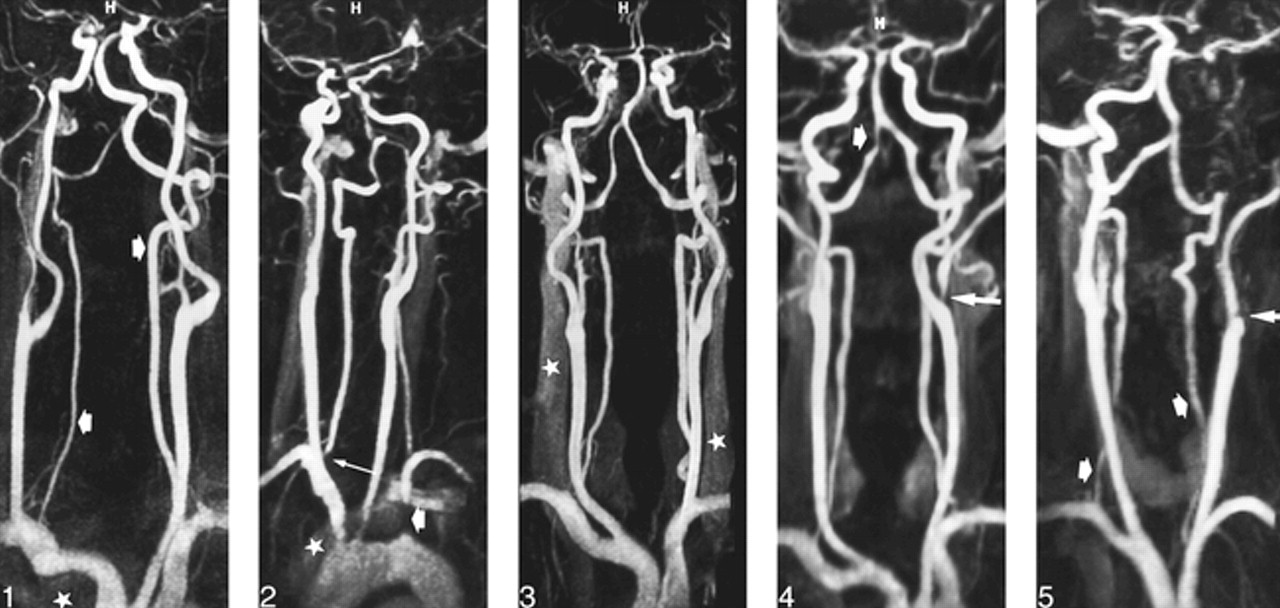
- fig 1.
46-year-old man referred for an ischemic event in the posterior fossa. Frontal view from contrast-enhanced MR angiogram (4.6/1.8/1, voxel size = 0.95 × 0.76 × 0.82) shows accurate delineation of vertebrobasilar system (arrows), despite wrap-around artifacts (star) at lower part of imaging volume that slightly degrade image quality.
fig 2. 75-year-old man referred for a subclavian steal syndrome with occlusion of initial portion of left subclavian artery (short arrow) and stenosis at origin of right vertebral artery (long arrow). Oblique view from MR angiogram (4.6/1.8/1, voxel size = 0.95 × 0.76 × 0.82) shows a rapid fall-off of signal intensity at inferior part of imaging volume and a moderate enhancement of left brachiocephalic vein (star), which degrades image quality at level of aortic arch and at origin of great vessels.
fig 3. 65-year-old man with atherosclerotic disease. Frontal view from MR angiogram (4.6/1.8/1, voxel size = 0.95 × 0.76 × 0.82) shows accurate delineation of cervical arteries, despite a moderate enhancement of jugular veins (stars).
fig 4. 56-year-old man with left external carotid stenosis (long arrow) and severe short stenosis at the distal portion of the right vertebral artery (short arrow). Frontal view from MR angiogram (4.6/1.8/1, voxel size = 1.3 × 1.29 × 1.25) shows a fair delineation of artery outlines despite a slight blurring of arterial lumen.
fig 5. 43-year-old woman with occlusive dissection of left internal carotid artery (long arrow). MR angiogram (4.6/1.8/1, voxel size = 1.3 × 1.29 × 1.25) shows a poor delineation of proximal segment of vertebral arteries (short arrows).

Tables
 TABLE 1:
Comparison of parameters of the two gadolinium-enhanced MR angiographic sequences
- TABLE 2:
Distribution of image artifacts of contrast-enhanced MR angiograms according to the voxel size
- TABLE 3:
Grading scale of contrast-enhanced MR angiograms according to the voxel size
- TABLE 4:
Findings of SNR (mean value, SD) measured in three ROIs according to the voxel size of MR angiograms
In this issue


Jump to section
Related Articles
Cited By...
- CTA for Screening of Complicated Atherosclerotic Carotid Plaque--American Heart Association Type VI Lesions as Defined by MRI
- Quality-Evaluation Scheme for Cerebral Time-Resolved 3D Contrast-Enhanced MR Angiography Techniques
- 3D High-Spatial-Resolution Cerebral MR Venography at 3T: A Contrast-Dose-Reduction Study
- Noninvasive Detection of Steno-Occlusive Disease of the Supra-Aortic Arteries With Three-Dimensional Contrast-Enhanced Magnetic Resonance Angiography: A Prospective, Intra-Individual Comparative Analysis With Digital Subtraction Angiography