Advertisement

Main menu

  • Home
  • Content
    • Current Issue
    • Accepted Manuscripts
    • Article Preview
    • Past Issue Archive
    • Video Articles
    • AJNR Case Collection
    • Case of the Week Archive
    • Case of the Month Archive
    • Classic Case Archive
  • Special Collections
    • AJNR Awards
    • Low-Field MRI
    • Alzheimer Disease
    • ASNR Foundation Special Collection
    • Photon-Counting CT
    • View All
  • Multimedia
    • AJNR Podcasts
    • AJNR SCANtastic
    • Trainee Corner
    • MRI Safety Corner
    • Imaging Protocols
  • For Authors
    • Submit a Manuscript
    • Submit a Video Article
    • Submit an eLetter to the Editor/Response
    • Manuscript Submission Guidelines
    • Statistical Tips
    • Fast Publishing of Accepted Manuscripts
    • Graphical Abstract Preparation
    • Imaging Protocol Submission
    • Author Policies
  • About Us
    • About AJNR
    • Editorial Board
    • Editorial Board Alumni
  • More
    • Become a Reviewer/Academy of Reviewers
    • Subscribers
    • Permissions
    • Alerts
    • Feedback
    • Advertisers
    • ASNR Home

User menu

  • Alerts
  • Log in

Search

  • Advanced search
American Journal of Neuroradiology

American Journal of Neuroradiology

ASHNR American Society of Functional Neuroradiology ASHNR American Society of Pediatric Neuroradiology ASSR
  • Alerts
  • Log in

Advanced Search

  • Home
  • Content
    • Current Issue
    • Accepted Manuscripts
    • Article Preview
    • Past Issue Archive
    • Video Articles
    • AJNR Case Collection
    • Case of the Week Archive
    • Case of the Month Archive
    • Classic Case Archive
  • Special Collections
    • AJNR Awards
    • Low-Field MRI
    • Alzheimer Disease
    • ASNR Foundation Special Collection
    • Photon-Counting CT
    • View All
  • Multimedia
    • AJNR Podcasts
    • AJNR SCANtastic
    • Trainee Corner
    • MRI Safety Corner
    • Imaging Protocols
  • For Authors
    • Submit a Manuscript
    • Submit a Video Article
    • Submit an eLetter to the Editor/Response
    • Manuscript Submission Guidelines
    • Statistical Tips
    • Fast Publishing of Accepted Manuscripts
    • Graphical Abstract Preparation
    • Imaging Protocol Submission
    • Author Policies
  • About Us
    • About AJNR
    • Editorial Board
    • Editorial Board Alumni
  • More
    • Become a Reviewer/Academy of Reviewers
    • Subscribers
    • Permissions
    • Alerts
    • Feedback
    • Advertisers
    • ASNR Home
  • Follow AJNR on Twitter
  • Visit AJNR on Facebook
  • Follow AJNR on Instagram
  • Join AJNR on LinkedIn
  • RSS Feeds

AJNR Awards, New Junior Editors, and more. Read the latest AJNR updates

Research ArticleSpine Imaging and Spine Image-Guided Interventions

Spinal Intradural Capillary Hemangioma: MR Findings

Ji Hoon Shin, Ho Kyu Lee, Sang Ryong Jeon and Sung Hye Park
American Journal of Neuroradiology May 2000, 21 (5) 954-956;
Ji Hoon Shin
  • Find this author on Google Scholar
  • Find this author on PubMed
  • Search for this author on this site
Ho Kyu Lee
  • Find this author on Google Scholar
  • Find this author on PubMed
  • Search for this author on this site
Sang Ryong Jeon
  • Find this author on Google Scholar
  • Find this author on PubMed
  • Search for this author on this site
Sung Hye Park
  • Find this author on Google Scholar
  • Find this author on PubMed
  • Search for this author on this site
  • Article
  • Figures & Data
  • Info & Metrics
  • Responses
  • References
  • PDF
Loading

Abstract

Summary: We report a case of a spinal intradural capillary hemangioma. On MR images, a well-circumscribed intradural mass was detected at the T8–T9 level. The signal intensity of the mass relative to the spinal cord was isointense on T1-weighted images, hyperintense on T2-weighted images, and showed homogeneous, strong enhancement on contrast-enhanced T1-weighted images. The mass had both extramedullary and intramedullary components.

Capillary hemangiomas are typically superficial lesions found in the skin or mucosa of the head and neck; intradural locations are rare (1–4). We report a case of an intradural capillary hemangioma in the thoracic spine.

Case Report

A 66-year-old woman initially presented with an 8-month history of lower back pain. A month before admission, she had begun to use a wheelchair owing to weakness of the lower extremities. Preoperative neurologic examination revealed grade 3 paraparesis and sensory abnormality.

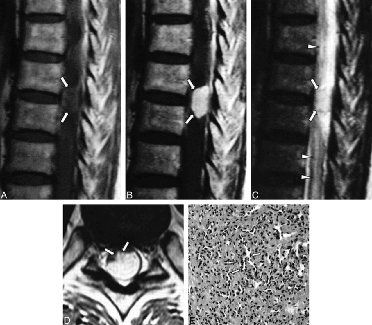
MR studies of the thoracic spine showed a well-defined intradural mass at the T8–T9 level with a diameter of 1.3 cm and a length of 2 cm. The signal intensity of the mass relative to the spinal cord was isointense on T1-weighted images, hyperintense on T2-weighted images (Fig 1A and C), and showed homogeneous, strong enhancement on contrast-enhanced T1-weighted images (Fig 1B and D). The spinal cord was compressed and displaced anteriorly and to the right (Fig 1D), which caused diffuse cord edema from T4 to the level of the conus medullaris (Fig 1C). The mass had an intramedullary component in the posterior aspect of the spinal cord (Fig 1D).

fig 1.
  • Download figure
  • Open in new tab
  • Download powerpoint
fig 1.

66-year-old woman with lower back pain.

A, T1-weighted (550/12 [TR/TE]) sagittal MR image shows a well-defined, oval mass at the dorsal aspect of the T8–T9 level (arrows). The signal intensity of the mass is isointense with the spinal cord.

B, Contrast-enhanced T1-weighted (550/12) sagittal MR image shows homogenous, intense enhancement of the mass (arrows).

C, On T2-weighted (3600/112) sagittal MR image, the signal intensity is hyperintense relative to the spinal cord (arrows). Note the associated diffuse cord edema (arrowheads).

D, On contrast-enhanced T1-weighted (874/12) axial MR image, the spinal cord is markedly compressed by the posterolaterally located mass (arrows).

E, Low-power photomicrograph shows the tumor to be composed of variably sized but relatively small capillaries (asterisks), pericytes (arrows), and some stromal cells (fibroblasts) (arrowheads) (hematoxylin-eosin, original magnification ×100).

At laminectomy of the T8–T9 vertebrae, a reddish mass was seen upon resection of the dura. The mass, which was easily separated from the dura, had a broad base with the spinal cord, and adhered to it. Removal of the mass was incomplete, owing to significant bleeding that occurred during the operation, and a duraplasty was performed after the mass was removed. The surgeon thought the mass was primarily subpial, as it grew exophytically into the subarachnoid space.

Microscopically, the lesion was composed of capillary-sized blood vessels and a little stroma; there were no foamy stromal cells, as seen in hemangioblastomas (Fig 1E). Factor VIII-related antigen, which is a marker for endothelial cells, was strongly immunoreactive in the lining cells of the capillaries. Smooth muscle actin immunoreactive cells surrounding the capillaries were considered to be pericytes. The diagnosis was capillary hemangioma.

After surgery, the lower extremity weakness and the lower back pain improved markedly, and the patient was able to walk unassisted. A follow-up MR examination 6 months later showed no definite residual tumor, and there was resolution of the previous cord edema.

Discussion

Hemangiomas are common soft-tissue tumors, especially in children (5). They are seen in cutaneous and subcutaneous locations but have rarely been encountered in the neuraxis (1–4, 6–8). Histologically, hemangiomas are divided into capillary and cavernous types, depending on the dominant vessel size at microscopy (4, 5). Capillary hemangiomas are the more common form and consist of capillary-sized vessels lined with flattened endothelium separated by a collagenous stroma. They usually appear in childhood, but some cases are found in adults. Within the neuraxis, cases of capillary hemangiomas in the meninges, cauda equina, epidural, or intramedullary locations are rare (1–4, 6, 9). In the intradural spine, only a few cases of capillary hemangioma of the cauda equina or of a purely intramedullary location have been reported (1–4).

The MR findings in this case are similar to those in previously reported capillary hemangiomas at other sites (5, 6, 9). That is, the signal intensity is isointense with the spinal cord on T1-weighted images, hyperintense relative to the spinal cord on T2-weighted images, and shows homogeneous, strong enhancement on contrast-enhanced T1-weighted images. Because the tumor in this patient was primarily intradural extramedullary, the differential diagnosis with other intradural extramedullary tumors was important. These signal characteristics could help differentiate this type of tumor from meningioma, a much more common intradural extramedullary tumor, as meningiomas display low or isointense signal intensity on T2-weighted images. Because the signal characteristics of the intradural hemangioma are similar to those of intradural extramedullary neurogenic tumors, such as schwannoma, differentiating between the two is difficult.

The anteriorly displaced spinal cord and adhesions of the mass to the posterolateral aspect of the spinal cord were observed during surgery. We speculated that this capillary hemangioma originated from penetrating pial plexus of the posterior spinal artery in the superficial portion of the spinal cord and had grown exophytically into the intradural extramedullary subarachnoid space. Although the natural history of capillary hemangiomas has not been well described, there is a significant risk of bleeding and enlargement, leading to neurologic deterioration. The current treatment of an intradural hemangioma consists of total excision (7).

Acknowledgments

We thank Dae Chul Suh, Choong Gon Choi, and Sung Chul Rhim for their continuous support.

Footnotes

  • ↵1 Address reprint requests to Ho Kyu Lee, MD, Department of Diagnostic Radiology, Asan Medical Center, 388-1 Poongnap-Dong, Songpa-Ku, Seoul, 138-736, South Korea.

References

  1. ↵
    Hanakita J, Suwa H, Nagayasu S, Suzuki H. Capillary hemangioma in the cauda equina: neuroradiological findings. Neuroradiology 1991;33:458-461
    PubMed
  2. Mawk JR, Leibrock LG, McComb RD, Trembath EJ. Metameric capillary hemangioma producing complete myelographic block in an infant. J Neurosurg 1987;67:456-459
    PubMed
  3. Holtzman RN, Brisson PM, Pearl RE, Gruber ML. Lobular capillary hemangioma of the cauda equina: case report. J Neurosurg 1999;90:239-241
    PubMed
  4. ↵
    Hida K, Tada M, Iwasaki Y, Abe H. Intramedullary disseminated capillary hemangioma with localized spinal cord swelling: case report. Neurosurgery 1993;33:1099-1101
    PubMed
  5. ↵
    Dillon WP, Som PM, Rosenau W. Hemangioma of the nasal vault: MR and CT features. Radiology 1991;180:761-765
    PubMed
  6. Gupta S, Kumar S, Banerji D, Pandey R, Gujral R. Magnetic resonance imaging features of an epidural spinal haemangioma. Australas Radiol 1996;40:342-344
    PubMed
  7. ↵
    Mastronardi L, Ferrante L, Scarpinati M, Gagliardi FM, Celli P, Fortuna A. Intradural extramedullary cavernous angioma: case report. Neurosurgery 1991;29:924-926
    PubMed
  8. Heimberger K, Schnaberth G, Koos W, Pendl G, Auff E. Spinal cavernous haemangioma (intradural-extramedullary) underlying repeated subarachnoid haemorrhage. J Neurol 1982;226:289-293
    PubMed
  9. Willing SJ, Faye-Petersen O, Aronin P, Faith S. Radiologic-pathologic correlation: capillary hemangioma of the meninges. AJNR Am J Neuroradiol 1993;14:529-536
    Abstract/FREE Full Text
  • Received August 30, 1999.
  • Copyright © American Society of Neuroradiology
View Abstract
PreviousNext
Back to top

In this issue

American Journal of Neuroradiology
Vol. 21, Issue 5
1 May 2000
  • Table of Contents
  • Index by author
Advertisement
Print
Download PDF
Email Article

Thank you for your interest in spreading the word on American Journal of Neuroradiology.

NOTE: We only request your email address so that the person you are recommending the page to knows that you wanted them to see it, and that it is not junk mail. We do not capture any email address.

Enter multiple addresses on separate lines or separate them with commas.
Spinal Intradural Capillary Hemangioma: MR Findings
(Your Name) has sent you a message from American Journal of Neuroradiology
(Your Name) thought you would like to see the American Journal of Neuroradiology web site.
CAPTCHA
This question is for testing whether or not you are a human visitor and to prevent automated spam submissions.
Cite this article
Ji Hoon Shin, Ho Kyu Lee, Sang Ryong Jeon, Sung Hye Park
Spinal Intradural Capillary Hemangioma: MR Findings
American Journal of Neuroradiology May 2000, 21 (5) 954-956;

Citation Manager Formats

  • BibTeX
  • Bookends
  • EasyBib
  • EndNote (tagged)
  • EndNote 8 (xml)
  • Medlars
  • Mendeley
  • Papers
  • RefWorks Tagged
  • Ref Manager
  • RIS
  • Zotero
0 Responses
Respond to this article
Share
Bookmark this article
Spinal Intradural Capillary Hemangioma: MR Findings
Ji Hoon Shin, Ho Kyu Lee, Sang Ryong Jeon, Sung Hye Park
American Journal of Neuroradiology May 2000, 21 (5) 954-956;
del.icio.us logo Twitter logo Facebook logo Mendeley logo
  • Tweet Widget
  • Facebook Like
  • Google Plus One

Jump to section

  • Article
    • Abstract
    • Case Report
    • Discussion
    • Acknowledgments
    • Footnotes
    • References
  • Figures & Data
  • Info & Metrics
  • Responses
  • References
  • PDF

Related Articles

  • No related articles found.
  • PubMed
  • Google Scholar

Cited By...

  • Mystery Case: A 78-year-old man with a gait disorder
  • Spinal Intradural Extramedullary Capillary Hemangioma: MR Imaging Findings
  • Crossref
  • Google Scholar

This article has not yet been cited by articles in journals that are participating in Crossref Cited-by Linking.

More in this TOC Section

  • Characteristics of SIH Type I Culprit Lesions
  • Management Outcomes For VO Spine Biopsy
  • Cone Beam CT Myelography
Show more SPINE IMAGING AND SPINE IMAGE-GUIDED INTERVENTIONS

Similar Articles

Advertisement

Indexed Content

  • Current Issue
  • Accepted Manuscripts
  • Article Preview
  • Past Issues
  • Editorials
  • Editor's Choice
  • Fellows' Journal Club
  • Letters to the Editor
  • Video Articles

Cases

  • Case Collection
  • Archive - Case of the Week
  • Archive - Case of the Month
  • Archive - Classic Case

More from AJNR

  • Trainee Corner
  • Imaging Protocols
  • MRI Safety Corner
  • Book Reviews

Multimedia

  • AJNR Podcasts
  • AJNR Scantastics

Resources

  • Turnaround Time
  • Submit a Manuscript
  • Submit a Video Article
  • Submit an eLetter to the Editor/Response
  • Manuscript Submission Guidelines
  • Statistical Tips
  • Fast Publishing of Accepted Manuscripts
  • Graphical Abstract Preparation
  • Imaging Protocol Submission
  • Evidence-Based Medicine Level Guide
  • Publishing Checklists
  • Author Policies
  • Become a Reviewer/Academy of Reviewers
  • News and Updates

About Us

  • About AJNR
  • Editorial Board
  • Editorial Board Alumni
  • Alerts
  • Permissions
  • Not an AJNR Subscriber? Join Now
  • Advertise with Us
  • Librarian Resources
  • Feedback
  • Terms and Conditions
  • AJNR Editorial Board Alumni

American Society of Neuroradiology

  • Not an ASNR Member? Join Now

© 2025 by the American Society of Neuroradiology All rights, including for text and data mining, AI training, and similar technologies, are reserved.
Print ISSN: 0195-6108 Online ISSN: 1936-959X

Powered by HighWire