
Article Figures & Data
Figures
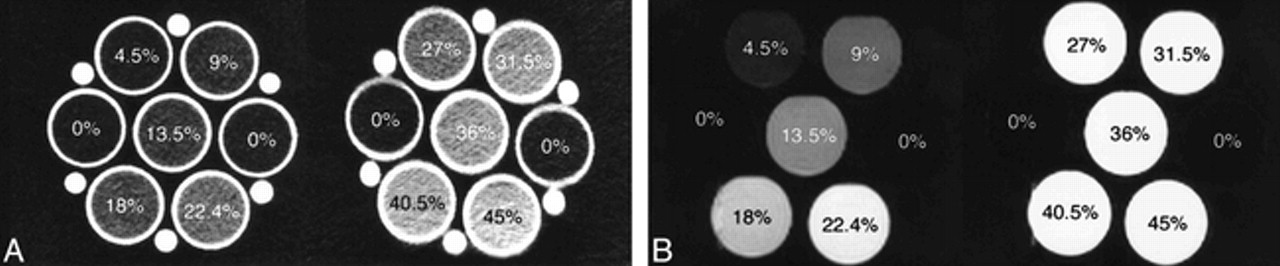
- FIG 1.
CSF (Hct, 0%) and each simulated SAH (ranging from Hct 4.5% to 45%).
A, CT scan.
B, Turbo-FLAIR image (9000/119 [TR/effective TE]; inversion time, 2200 ms).
- FIG 2.
Plots of simulated SAH:CSF ratio versus Hct (%)
- FIG 3.
Plots of CT value (in HU) versus Hct (%) and correlation with the average CT value of the normal cortical gray matter
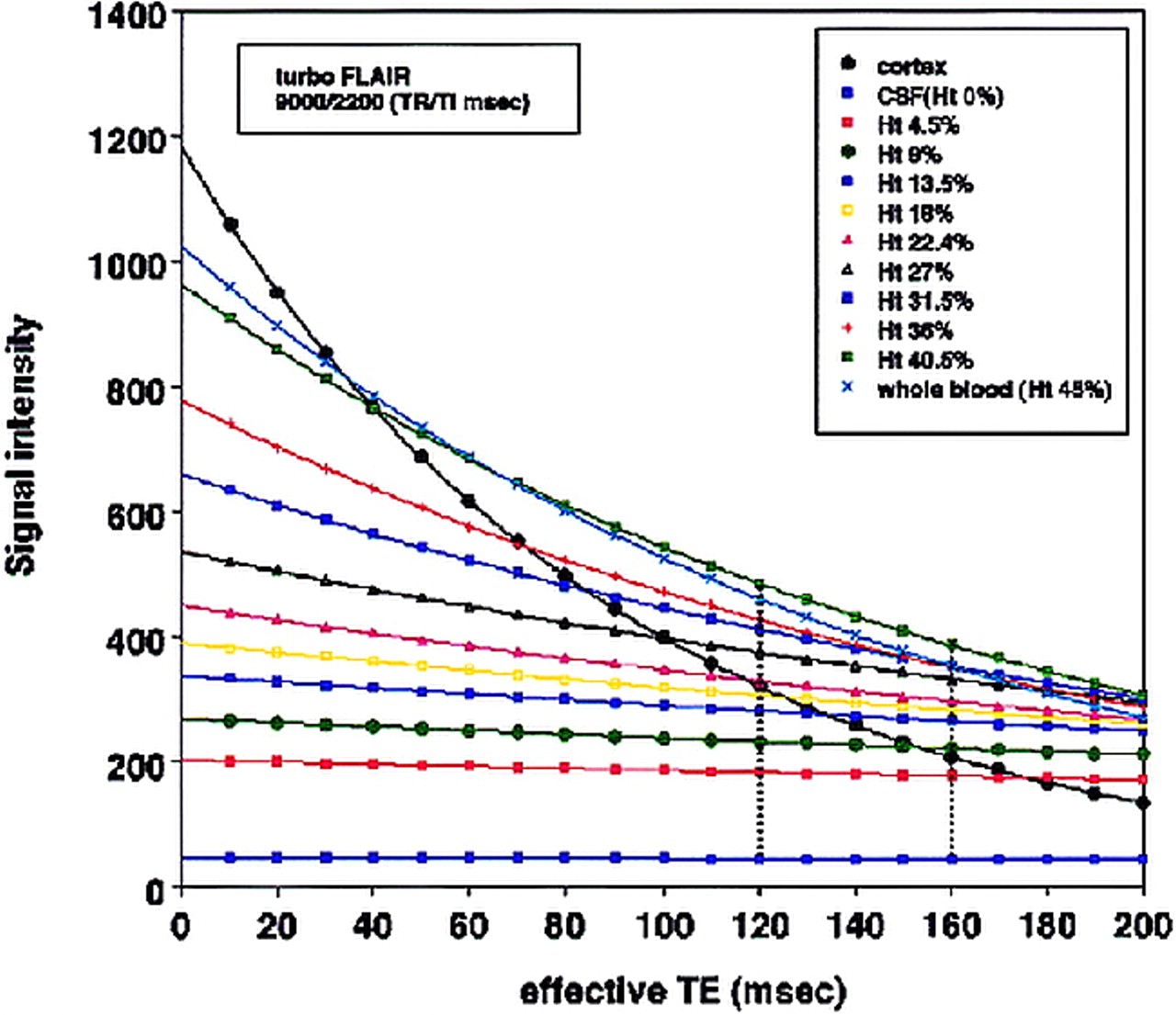
- FIG 4.
T2 relaxation curves of normal cortical gray matter, CSF (Hct, 0%), and each simulated SAH (ranging from Hct 4.5% to 45%) as a function of effective TE for a specific inversion time (2200 ms) and TR (9000) in a turbo-FLAIR sequence by using a theoretical equation for turbo inversion recovery signal intensity


