Advertisement

Main menu

  • Home
  • Content
    • Current Issue
    • Accepted Manuscripts
    • Article Preview
    • Past Issue Archive
    • Video Articles
    • AJNR Case Collection
    • Case of the Week Archive
    • Case of the Month Archive
    • Classic Case Archive
  • Special Collections
    • AJNR Awards
    • Low-Field MRI
    • Alzheimer Disease
    • ASNR Foundation Special Collection
    • Photon-Counting CT
    • View All
  • Multimedia
    • AJNR Podcasts
    • AJNR SCANtastic
    • Trainee Corner
    • MRI Safety Corner
    • Imaging Protocols
  • For Authors
    • Submit a Manuscript
    • Submit a Video Article
    • Submit an eLetter to the Editor/Response
    • Manuscript Submission Guidelines
    • Statistical Tips
    • Fast Publishing of Accepted Manuscripts
    • Graphical Abstract Preparation
    • Imaging Protocol Submission
    • Author Policies
  • About Us
    • About AJNR
    • Editorial Board
    • Editorial Board Alumni
  • More
    • Become a Reviewer/Academy of Reviewers
    • Subscribers
    • Permissions
    • Alerts
    • Feedback
    • Advertisers
    • ASNR Home

User menu

  • Alerts
  • Log in

Search

  • Advanced search
American Journal of Neuroradiology

American Journal of Neuroradiology

ASHNR American Society of Functional Neuroradiology ASHNR American Society of Pediatric Neuroradiology ASSR
  • Alerts
  • Log in

Advanced Search

  • Home
  • Content
    • Current Issue
    • Accepted Manuscripts
    • Article Preview
    • Past Issue Archive
    • Video Articles
    • AJNR Case Collection
    • Case of the Week Archive
    • Case of the Month Archive
    • Classic Case Archive
  • Special Collections
    • AJNR Awards
    • Low-Field MRI
    • Alzheimer Disease
    • ASNR Foundation Special Collection
    • Photon-Counting CT
    • View All
  • Multimedia
    • AJNR Podcasts
    • AJNR SCANtastic
    • Trainee Corner
    • MRI Safety Corner
    • Imaging Protocols
  • For Authors
    • Submit a Manuscript
    • Submit a Video Article
    • Submit an eLetter to the Editor/Response
    • Manuscript Submission Guidelines
    • Statistical Tips
    • Fast Publishing of Accepted Manuscripts
    • Graphical Abstract Preparation
    • Imaging Protocol Submission
    • Author Policies
  • About Us
    • About AJNR
    • Editorial Board
    • Editorial Board Alumni
  • More
    • Become a Reviewer/Academy of Reviewers
    • Subscribers
    • Permissions
    • Alerts
    • Feedback
    • Advertisers
    • ASNR Home
  • Follow AJNR on Twitter
  • Visit AJNR on Facebook
  • Follow AJNR on Instagram
  • Join AJNR on LinkedIn
  • RSS Feeds

AJNR Awards, New Junior Editors, and more. Read the latest AJNR updates

Research ArticleHead and Neck Imaging

Imaging of Nonlaryngeal Neuroendocrine Carcinoma

Uday S. Kanamalla, Prabhakar P. Kesava and H. Stan McGuff
American Journal of Neuroradiology April 2000, 21 (4) 775-778;
Uday S. Kanamalla
aFrom the Section of Neuroradiology (U.S.K., P.P.K.) and Department of Pathology (H.S.M.), University of Texas Health Science Center, San Antonio, TX.
  • Find this author on Google Scholar
  • Find this author on PubMed
  • Search for this author on this site
Prabhakar P. Kesava
aFrom the Section of Neuroradiology (U.S.K., P.P.K.) and Department of Pathology (H.S.M.), University of Texas Health Science Center, San Antonio, TX.
  • Find this author on Google Scholar
  • Find this author on PubMed
  • Search for this author on this site
H. Stan McGuff
aFrom the Section of Neuroradiology (U.S.K., P.P.K.) and Department of Pathology (H.S.M.), University of Texas Health Science Center, San Antonio, TX.
  • Find this author on Google Scholar
  • Find this author on PubMed
  • Search for this author on this site
  • Article
  • Figures & Data
  • Info & Metrics
  • Responses
  • References
  • PDF
Loading

Article Figures & Data

Figures

  • fig 1.
    • Download figure
    • Open in new tab
    • Download powerpoint
    fig 1.

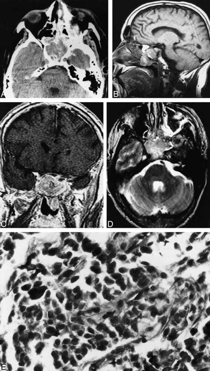
    Case 1. A, Axial unenhanced CT scan shows a soft-tissue density mass in the sphenoid sinus. The mass is seen to expand the sinus.

    B, Sagittal T1-weighted MR image (600/16/1 [TR/TE/excitations]) shows the nasopharyngeal component of the sphenoid mass. Note that the floor of the sella appears intact.

    C, Coronal contrast-enhanced T1-weighted MR image (433/17/1) shows mild heterogeneous enhancement of the sphenoid sinus mass.

    D, Axial fast spin-echo T2-weighted MR image (3250/95/3) shows the sphenoid sinus mass to be heterogeneously hyperintense.

    E, Neuroendocrine carcinoma of the sphenoid sinus consisting of small pleomorphic cells exhibiting crowded hyperchromatic nuclei (hematoxylin and eosin stain; original magnification, ×40).

  • fig 2.
    • Download figure
    • Open in new tab
    • Download powerpoint
    fig 2.

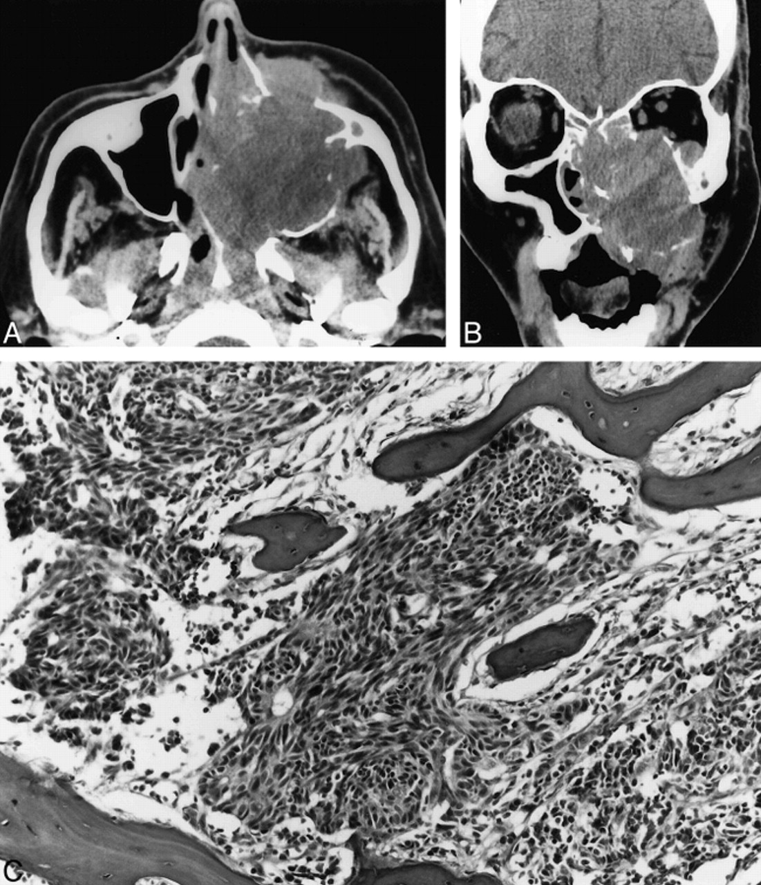
    Case 2. A, Axial unenhanced CT scan shows a large expansile maxillary sinus mass with extension into the nasal cavity, the nasopharynx, the infratemporal fossa, and the subcutaneous tissues of the left maxillary region.

    B, Coronal unenhanced CT scan shows a large expansile mass in the left maxillary sinus that erodes the sinus walls and extends into the nasal cavity, left orbit, and subcutaneous soft tissues of the maxillary region.

    C, Poorly differentiated neuroendocrine carcinoma infiltrating maxillary bone. The tumor consists of small pleomorphic spindled cells with hyperchromatic nuclei. Focal rosette formation is noted (hematoxylin and eosin stain; original magnification, ×10).

  • fig 3.
    • Download figure
    • Open in new tab
    • Download powerpoint
    fig 3.

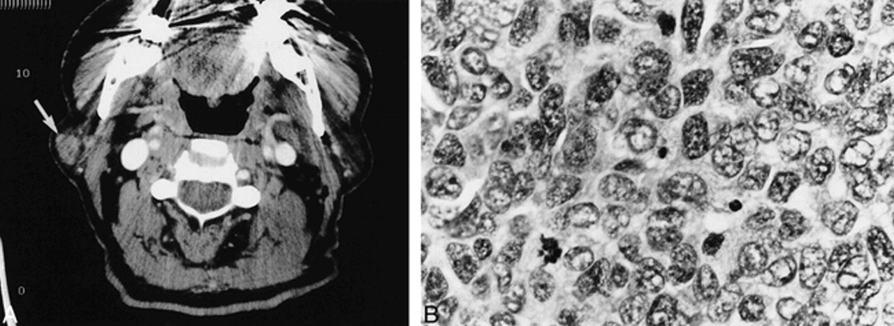
    Case 3. A, Axial contrast-enhanced CT scan shows a mildly enhancing circumscribed mass (arrow) in the lateral aspect of the superficial lobe of the right parotid gland.

    B, Metastatic poorly differentiated neuroendocrine carcinoma to an intraparotid lymph node. The tumor cells display pleomorphic vesicular nuclei with finely dispersed chromatin, scant cytoplasm, and a high mitotic rate (hematoxylin and eosin stain; original magnification, ×40).

PreviousNext
Back to top

In this issue

American Journal of Neuroradiology
Vol. 21, Issue 4
1 Apr 2000
  • Table of Contents
  • Index by author
Advertisement
Print
Download PDF
Email Article

Thank you for your interest in spreading the word on American Journal of Neuroradiology.

NOTE: We only request your email address so that the person you are recommending the page to knows that you wanted them to see it, and that it is not junk mail. We do not capture any email address.

Enter multiple addresses on separate lines or separate them with commas.
Imaging of Nonlaryngeal Neuroendocrine Carcinoma
(Your Name) has sent you a message from American Journal of Neuroradiology
(Your Name) thought you would like to see the American Journal of Neuroradiology web site.
CAPTCHA
This question is for testing whether or not you are a human visitor and to prevent automated spam submissions.
Cite this article
Uday S. Kanamalla, Prabhakar P. Kesava, H. Stan McGuff
Imaging of Nonlaryngeal Neuroendocrine Carcinoma
American Journal of Neuroradiology Apr 2000, 21 (4) 775-778;

Citation Manager Formats

  • BibTeX
  • Bookends
  • EasyBib
  • EndNote (tagged)
  • EndNote 8 (xml)
  • Medlars
  • Mendeley
  • Papers
  • RefWorks Tagged
  • Ref Manager
  • RIS
  • Zotero
0 Responses
Respond to this article
Share
Bookmark this article
Imaging of Nonlaryngeal Neuroendocrine Carcinoma
Uday S. Kanamalla, Prabhakar P. Kesava, H. Stan McGuff
American Journal of Neuroradiology Apr 2000, 21 (4) 775-778;
del.icio.us logo Twitter logo Facebook logo Mendeley logo
  • Tweet Widget
  • Facebook Like
  • Google Plus One

Jump to section

  • Article
    • Abstract
    • Case Reports
    • Discussion
    • Conclusion
    • Footnotes
    • References
  • Figures & Data
  • Info & Metrics
  • Responses
  • References
  • PDF

Related Articles

  • No related articles found.
  • PubMed
  • Google Scholar

Cited By...

  • No citing articles found.
  • Crossref
  • Google Scholar

This article has not yet been cited by articles in journals that are participating in Crossref Cited-by Linking.

More in this TOC Section

  • FDG Uptake and Thyroid Cancer Post-BRAF Therapy
  • Normal Facial Nerve Enhancement on VIBE Sequence
  • Comprehensive Review of Inner Ear Anatomy on Photon-Counting CT
Show more Head and Neck Imaging

Similar Articles

Advertisement

Indexed Content

  • Current Issue
  • Accepted Manuscripts
  • Article Preview
  • Past Issues
  • Editorials
  • Editor's Choice
  • Fellows' Journal Club
  • Letters to the Editor
  • Video Articles

Cases

  • Case Collection
  • Archive - Case of the Week
  • Archive - Case of the Month
  • Archive - Classic Case

More from AJNR

  • Trainee Corner
  • Imaging Protocols
  • MRI Safety Corner

Multimedia

  • AJNR Podcasts
  • AJNR Scantastics

Resources

  • Turnaround Time
  • Submit a Manuscript
  • Submit a Video Article
  • Submit an eLetter to the Editor/Response
  • Manuscript Submission Guidelines
  • Statistical Tips
  • Fast Publishing of Accepted Manuscripts
  • Graphical Abstract Preparation
  • Imaging Protocol Submission
  • Evidence-Based Medicine Level Guide
  • Publishing Checklists
  • Author Policies
  • Become a Reviewer/Academy of Reviewers
  • News and Updates

About Us

  • About AJNR
  • Editorial Board
  • Editorial Board Alumni
  • Alerts
  • Permissions
  • Not an AJNR Subscriber? Join Now
  • Advertise with Us
  • Librarian Resources
  • Feedback
  • Terms and Conditions
  • AJNR Editorial Board Alumni

American Society of Neuroradiology

  • Not an ASNR Member? Join Now

© 2025 by the American Society of Neuroradiology All rights, including for text and data mining, AI training, and similar technologies, are reserved.
Print ISSN: 0195-6108 Online ISSN: 1936-959X

Powered by HighWire