
Article Figures & Data
Figures
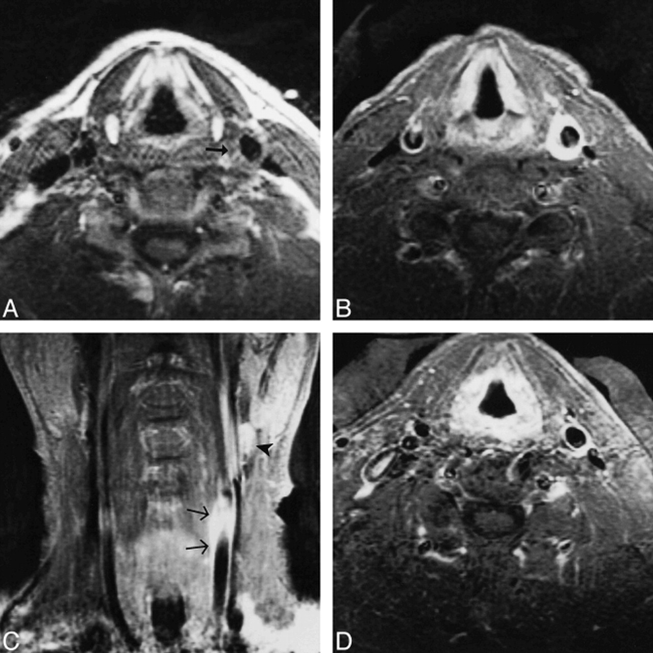
Fig 1. 48-year-old man with left-sided carotidynia.
A, Axial T1-weighted image (700/16/2 [TR/TE/excitations]) at the level of the distal common carotid artery. A skin marker has been placed over the area of swelling and tenderness. Note the abnormal soft-tissue signal surrounding the left carotid (arrow).
B, Axial contrast-enhanced T1-weighted image (550/16/2) shows striking enhancement of the tissue surrounding the artery.
C, Coronal contrast-enhanced T1-weighted image (550/16/2) shows the enhancement extending to the level of the carotid bifurcation (arrows). A lymph node is seen above the bifurcation (arrowhead).
D, Axial contrast-enhanced T1-weighted image (500/12/2) obtained several months later, while the patient remained asymptomatic, shows resolution of the thick rim of enhancement.
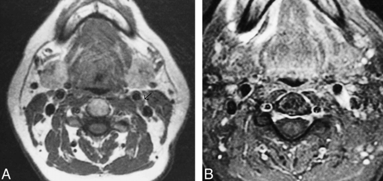
Fig 2. 31-year-old woman with right-sided carotidynia.
A, Axial T1-weighted image (700/11/2) at the level of the distal common carotid artery shows abnormal soft-tissue signal surrounding the carotid (arrows). The carotid itself is normal in contour and caliber.
B, Axial contrast-enhanced T1-weighted image at the same level shows marked enhancement of the tissue surrounding the vessel. Note the smooth margins of the enhancement as it extends toward the internal jugular vein, suggesting conformance to the fascial borders of the carotid sheath.
Fig 3. 51-year-old woman with left-sided carotidynia.
A, Axial T1-weighted image (560/15/2) at the level of the distal common carotid artery shows abnormal soft-tissue signal between the carotid and internal jugular vein (arrow).
B, Axial contrast-enhanced T1-weighted image (580/15/2) shows marked enhancement of the abnormal tissue. Note again how the smooth margins of the enhancement extend toward the internal jugular vein, in conformance with the expected borders of the carotid sheath.


Tables
 TABLE 1:
International Headache Society Classification Committee criteria for the diagnosis of idiopathic carotidynia


