
Article Figures & Data
Figures
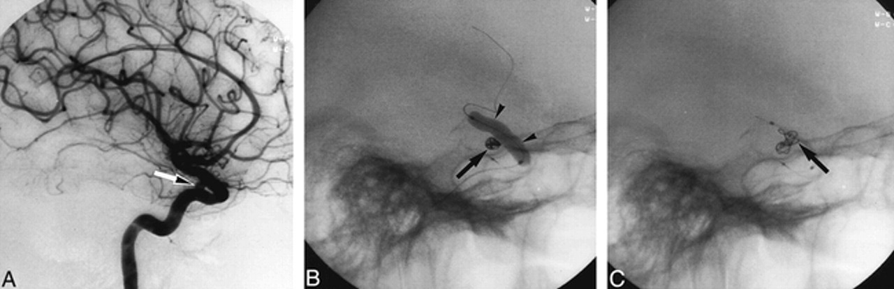
Fig 1. A, Selective angiogram of the left internal carotid artery. Small posterior paraclinoid aneurysm (arrow) with apparent small neck can be seen. B, During balloon-protected coiling, the GDC (arrow) fit well in the aneurysm. The inflated balloon (arrowheads) can be seen. C, After balloon deflation, the coil migrated out of the aneurysm (arrow) into the supraclinoid portion of the internal carotid artery
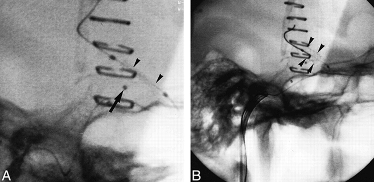
Fig 2. Before stent deployment (A), the tip of the Tracker double-tip microcatheter (arrow) was in the aneurysmal sac. The balloon-mounted stent (arrowheads) can be seen in the supraclinoid portion of the internal carotid artery. After stent deployment (B), the deployed stent (arrowheads) can be readily seen
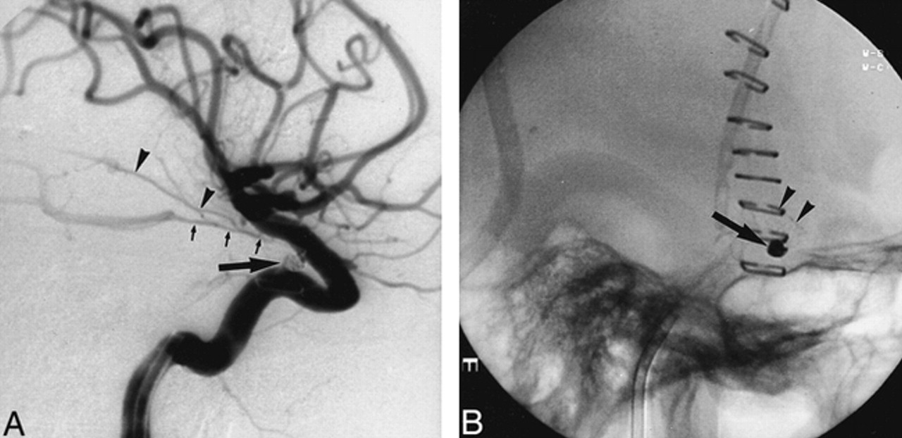
Fig 3. A, Aneurysm (arrow) is shown filled with coils. Note the patent posterior communicating artery (small arrow) and the anterior choroidal artery (arrowheads). B, Unsubtracted film shows the coils (arrow) and stent (arrowheads)
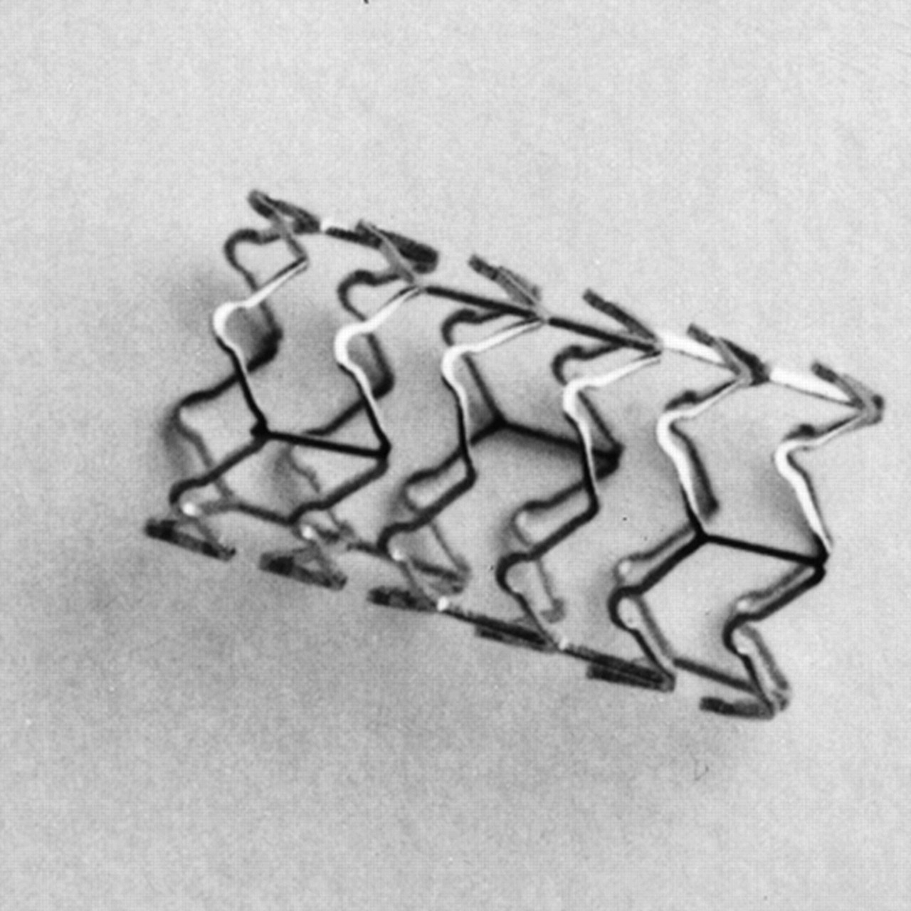
Fig 4. ACS Multilink RX Duet-stent. Note the design of the stent mesh, which allows optimal flexibility


