
Article Figures & Data
Figures
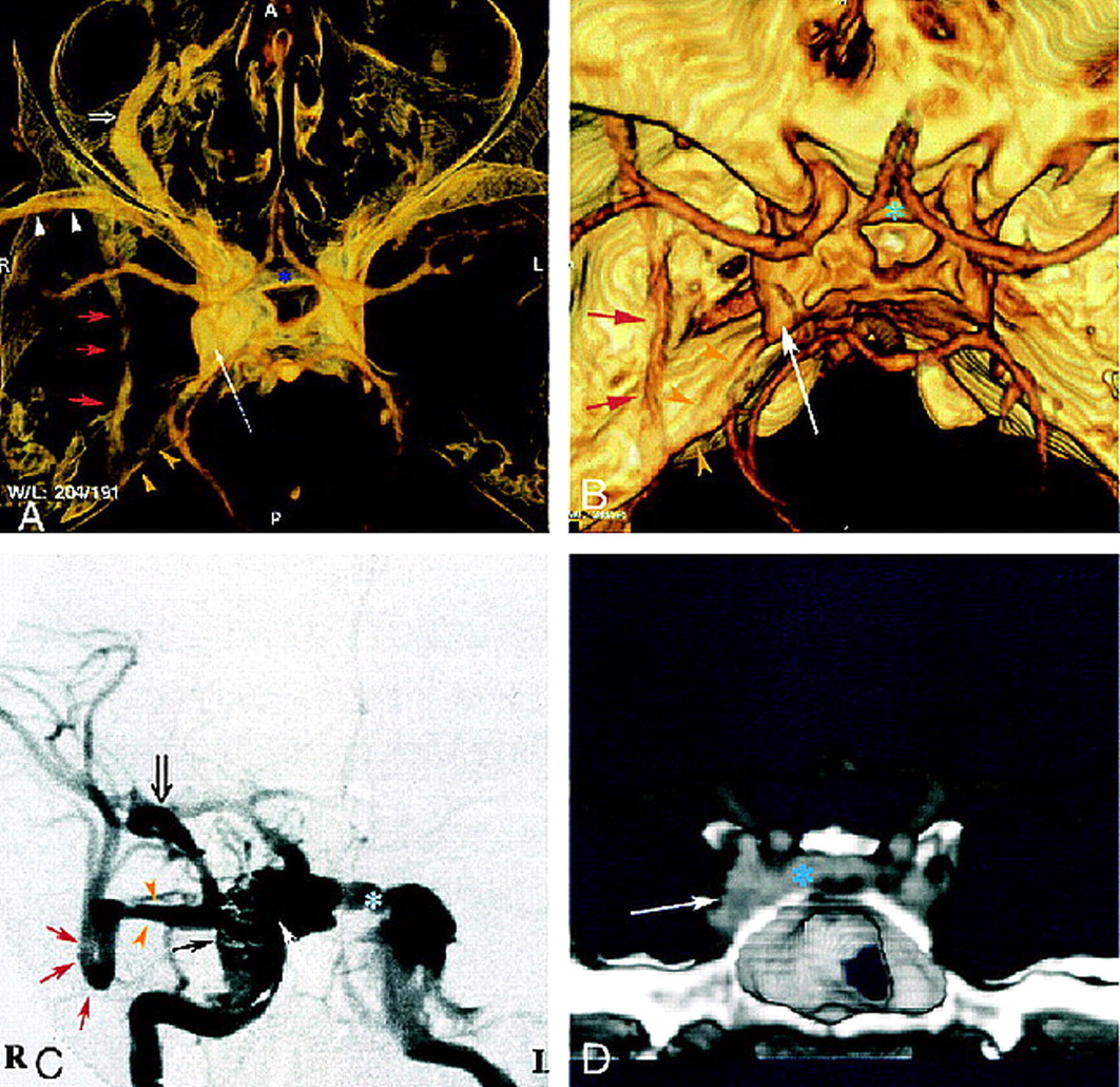
- fig 1.
Case 1: 40-year-old man with direct, posttraumatic, right-sided CCF.
A and B, Superior view of 3D CT angiogram obtained with the volume rendering technique shows an enlarged right cavernous sinus (long arrow ) with several draining veins: large right SOV (open arrow, A), anterior intercavernous sinus (asterisk ), inferior petrous sinus (yellow arrowheads), sphenoparietal sinus (white arrowheads, A), and paracavernous sinus (red arrows).
C, DSA during embolization with selective right internal carotid artery injection shows right cavernous fistula with very high flow and multiple venous drainage channels, including large right SOV (open arrow ), anterior intercavernous sinus (asterisk ), sphenoparietal sinus (yellow arrowheads), and paracavernous sinus (red arrows). Closed black arrow indicates GDC coils; long white arrow, right cavernous sinus.
D, Coronal view of 3D CT angiogram obtained with the volume rendering technique, using anterior cutting, clearly depicts intercavernous sinus (asterisk ). Arrow indicates right cavernous sinus.
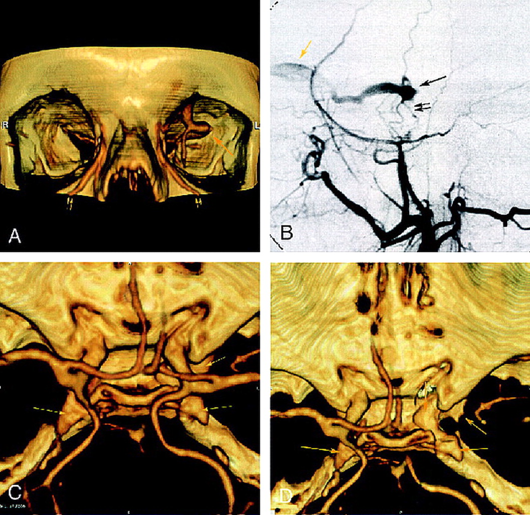
- fig 2.
Case 2: 69-year-old woman with dural cavernous fistula.
A, Frontal view of 3D CT angiogram obtained with the volume rendering technique shows an enlarged left SOV at the superior orbital fissure (single arrow ) and dilated angular veins (double arrows) bilaterally.
B, DSA (selective left external carotid injection, arterial phase, lateral view) shows rapid opacification of a portion of the cavernous sinus (single black arrow ) supplied by middle meningeal artery branches (double black arrows) and draining anteriorly into an enlarged left SOV (yellow arrow ).
C, 3D CT angiogram obtained with the volume rendering technique (superior view) shows bilateral enlarged cavernous sinuses (arrows).
D, CT angiogram (superior view) better depicts the prominent left cavernous sinus (arrows) after cutting to exclude supracavernous internal carotid artery.
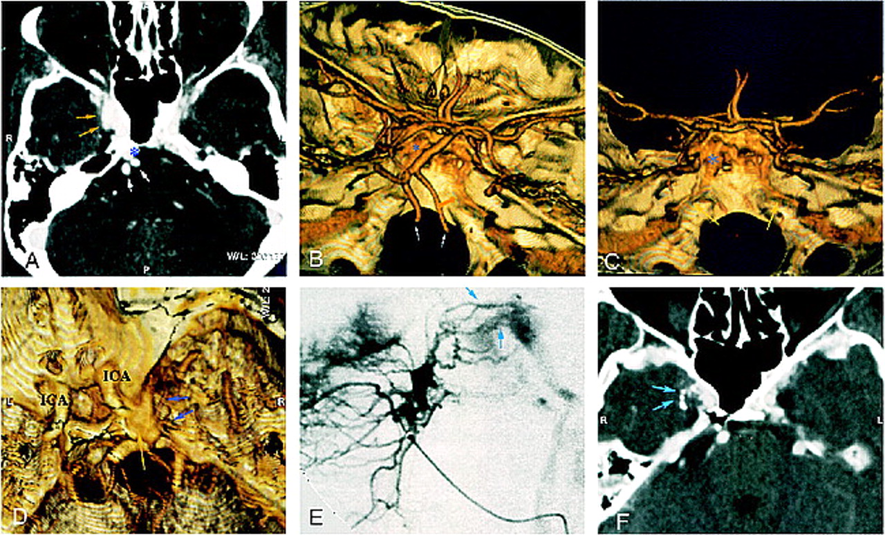
- fig 3.
Case 3: 54-year-old woman with dural CCF and right exophthalmos, chemosis, and dilated episcleral vessels.
A, CT angiogram (axial source image) shows an enlarged right cavernous sinus with irregular wall (yellow arrows) and large ipsilateral basilar plexus (asterisk ). White arrows indicate vertebral arteries.
B and C, Superolateral (B ) and posterior (C ) views of 3D CT angiogram obtained with the volume rendering technique show inferior petrosal sinuses along the posterior surface of the petrous bone (yellow arrows). Note the good delineation of skull and vascular anatomy. Asterisk indicates basilar plexus; white arrows, vertebral arteries.
D–F, Superolateral view of 3D CT angiogram obtained with the volume rendering technique (D ) shows enlarged cavernous sinus (yellow arrow ). Small vessels (blue arrows) might correspond to arteriovenous shunts, well depicted by lateral selective internal maxillary injection on DSA (E ). CT angiogram (axial source image) after superselective intraarterial embolization shows n-butyl cyanoacrylate in these arterial feeders (F ).
- fig 4.
Case 4: 69-year-old woman with a dural fistula involving bilateral cavernous sinuses.
A, CT angiogram (axial source image) shows enhancement of bilateral cavernous sinuses (double arrows) and enlarged left SOV (arrowhead ).
B, DSA (right common carotid injection, late arterial phase, anteroposterior view) shows early opacification of bilateral enlarged cavernous sinuses (arrows).
C, CT angiogram (axial source image), after partial embolization, shows partial thrombosis of left SOV (arrow ), without enhancement of left cavernous sinus (arrowheads), and persistent right enlarged cavernous sinus (asterisk ).
D, DSA (right common carotid injection, arterial phase, anteroposterior view) confirms rapid opacification of right cavernous sinus (asterisk ) and intercavernous sinus (thin arrow ), without enhancement of left cavernous sinus (thick arrow ).
- fig 5.
Schematic anatomic diagram of the venous vasculature of the skull base (superior view). 1, superior ophthalmic vein; 2, anterior intercavernous sinus; 3, inferior ophthalmic vein; 4, pterygoid plexus; 5, middle meningeal vein; 6, superior petrosal sinus; 7, inferior petrosal sinus; 8, basilar venous plexus; 9, transverse sinus; 10, posterior intercavernous sinus; 11, cavernous sinus; 12, sphenoparietal sinus


