
Article Figures & Data
Figures
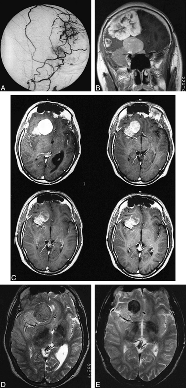
- fig 1.
Patient 1.
A, Superselective angiogram of the right external carotid artery (lateral view) shows blood supply of frontal and frontobasal meningiomas by branches of the frontal and parietal branches of the right middle meningeal artery.
B, Coronal T1-weighted spin-echo MR image (532/17/2) 12 hours after embolization shows persistent uptake of contrast medium in both meningiomas despite complete devascularization of the external carotid artery feeders.
C, Contrast-enhanced axial T1-weighted images (490/17/2) 12 hours (top left), 8 months (top right), 14 months (bottom left), and 22 months (bottom right) after embolization show a reduction in size and contrast enhancement of the meningioma. After 22 months, there is only a slight peripheral uptake of contrast medium.
D and E, Axial T2-weighted spin-echo images (1957/80/2) before (D) and 22 months after (E) embolization. The intensity changed from an isointense signal to a marked hypointense signal, which was probably due to intratumoral thrombosis.
- fig 2.
Progressive shrinkage of tumor size in patients 1 and 2 (tumor volume in cm3)
- fig 3.
Patient 2.
A, Coronal contrast-enhanced T1-weighted spin-echo MR image (490/17/2) before embolization and before antiedematous treatment shows a large right temporal meningioma with extensive midline shift.
B, Twelve hours after embolization there is a central necrosis and a slight reduction in size. Moreover, the medial portions of the tumor show diminished uptake of contrast medium.
C, Eight months after embolization there is marked reduction in tumor size. The central necrosis has completely disappeared and the medial portions of the tumor show a slight uptake of contrast medium.
D, Spectrum of the meningioma (single-voxel PRESS sequence, 1500/135/128, voxel size, 20 mm3) shows persistence of the increased Cho/Cr ratio and an absence of NAA. There is an alanine (Ala) doublet at 1.45 ppm and lipid (lip) signals at 0.8 to 1.2 ppm, which were not present before embolization, indicating intratumoral necrosis.


