Advertisement

Main menu

  • Home
  • Content
    • Current Issue
    • Accepted Manuscripts
    • Article Preview
    • Past Issue Archive
    • Video Articles
    • AJNR Case Collection
    • Case of the Week Archive
    • Case of the Month Archive
    • Classic Case Archive
  • Special Collections
    • AJNR Awards
    • Low-Field MRI
    • Alzheimer Disease
    • ASNR Foundation Special Collection
    • Photon-Counting CT
    • View All
  • Multimedia
    • AJNR Podcasts
    • AJNR SCANtastic
    • Trainee Corner
    • MRI Safety Corner
    • Imaging Protocols
  • For Authors
    • Submit a Manuscript
    • Submit a Video Article
    • Submit an eLetter to the Editor/Response
    • Manuscript Submission Guidelines
    • Statistical Tips
    • Fast Publishing of Accepted Manuscripts
    • Graphical Abstract Preparation
    • Imaging Protocol Submission
    • Author Policies
  • About Us
    • About AJNR
    • Editorial Board
    • Editorial Board Alumni
  • More
    • Become a Reviewer/Academy of Reviewers
    • Subscribers
    • Permissions
    • Alerts
    • Feedback
    • Advertisers
    • ASNR Home

User menu

  • Alerts
  • Log in

Search

  • Advanced search
American Journal of Neuroradiology

American Journal of Neuroradiology

ASHNR American Society of Functional Neuroradiology ASHNR American Society of Pediatric Neuroradiology ASSR
  • Alerts
  • Log in

Advanced Search

  • Home
  • Content
    • Current Issue
    • Accepted Manuscripts
    • Article Preview
    • Past Issue Archive
    • Video Articles
    • AJNR Case Collection
    • Case of the Week Archive
    • Case of the Month Archive
    • Classic Case Archive
  • Special Collections
    • AJNR Awards
    • Low-Field MRI
    • Alzheimer Disease
    • ASNR Foundation Special Collection
    • Photon-Counting CT
    • View All
  • Multimedia
    • AJNR Podcasts
    • AJNR SCANtastic
    • Trainee Corner
    • MRI Safety Corner
    • Imaging Protocols
  • For Authors
    • Submit a Manuscript
    • Submit a Video Article
    • Submit an eLetter to the Editor/Response
    • Manuscript Submission Guidelines
    • Statistical Tips
    • Fast Publishing of Accepted Manuscripts
    • Graphical Abstract Preparation
    • Imaging Protocol Submission
    • Author Policies
  • About Us
    • About AJNR
    • Editorial Board
    • Editorial Board Alumni
  • More
    • Become a Reviewer/Academy of Reviewers
    • Subscribers
    • Permissions
    • Alerts
    • Feedback
    • Advertisers
    • ASNR Home
  • Follow AJNR on Twitter
  • Visit AJNR on Facebook
  • Follow AJNR on Instagram
  • Join AJNR on LinkedIn
  • RSS Feeds

AJNR Awards, New Junior Editors, and more. Read the latest AJNR updates

Research ArticleSpine Imaging and Spine Image-Guided Interventions

Diffusion-Weighted MR Imaging of the Spinal Cord

Roland Bammer, Franz Fazekas, Michael Augustin, Josef Simbrunner, Siegrid Strasser-Fuchs, Thomas Seifert, Rudolf Stollberger and Hans-Peter Hartung
American Journal of Neuroradiology March 2000, 21 (3) 587-591;
Roland Bammer
aFrom the Department of Neurology (R.B., F.F., M.A., S.S.-F., T.S., H.-P.H.) and the Magnetic Resonance Institute (R.B., F.F., M.A., J.S., R.S.), Karl-Franzens University, Graz, Austria.
  • Find this author on Google Scholar
  • Find this author on PubMed
  • Search for this author on this site
Franz Fazekas
aFrom the Department of Neurology (R.B., F.F., M.A., S.S.-F., T.S., H.-P.H.) and the Magnetic Resonance Institute (R.B., F.F., M.A., J.S., R.S.), Karl-Franzens University, Graz, Austria.
  • Find this author on Google Scholar
  • Find this author on PubMed
  • Search for this author on this site
Michael Augustin
aFrom the Department of Neurology (R.B., F.F., M.A., S.S.-F., T.S., H.-P.H.) and the Magnetic Resonance Institute (R.B., F.F., M.A., J.S., R.S.), Karl-Franzens University, Graz, Austria.
  • Find this author on Google Scholar
  • Find this author on PubMed
  • Search for this author on this site
Josef Simbrunner
aFrom the Department of Neurology (R.B., F.F., M.A., S.S.-F., T.S., H.-P.H.) and the Magnetic Resonance Institute (R.B., F.F., M.A., J.S., R.S.), Karl-Franzens University, Graz, Austria.
  • Find this author on Google Scholar
  • Find this author on PubMed
  • Search for this author on this site
Siegrid Strasser-Fuchs
aFrom the Department of Neurology (R.B., F.F., M.A., S.S.-F., T.S., H.-P.H.) and the Magnetic Resonance Institute (R.B., F.F., M.A., J.S., R.S.), Karl-Franzens University, Graz, Austria.
  • Find this author on Google Scholar
  • Find this author on PubMed
  • Search for this author on this site
Thomas Seifert
aFrom the Department of Neurology (R.B., F.F., M.A., S.S.-F., T.S., H.-P.H.) and the Magnetic Resonance Institute (R.B., F.F., M.A., J.S., R.S.), Karl-Franzens University, Graz, Austria.
  • Find this author on Google Scholar
  • Find this author on PubMed
  • Search for this author on this site
Rudolf Stollberger
aFrom the Department of Neurology (R.B., F.F., M.A., S.S.-F., T.S., H.-P.H.) and the Magnetic Resonance Institute (R.B., F.F., M.A., J.S., R.S.), Karl-Franzens University, Graz, Austria.
  • Find this author on Google Scholar
  • Find this author on PubMed
  • Search for this author on this site
Hans-Peter Hartung
aFrom the Department of Neurology (R.B., F.F., M.A., S.S.-F., T.S., H.-P.H.) and the Magnetic Resonance Institute (R.B., F.F., M.A., J.S., R.S.), Karl-Franzens University, Graz, Austria.
  • Find this author on Google Scholar
  • Find this author on PubMed
  • Search for this author on this site
  • Article
  • Figures & Data
  • Info & Metrics
  • Responses
  • References
  • PDF
Loading

Article Figures & Data

Figures

  • fig 1.
    • Download figure
    • Open in new tab
    • Download powerpoint
    fig 1.

    Diffusion-weighted images obtained from volunteers in three mutually orthogonal directions.

    Top row, Direction-dependent diffusion-weighted interleaved echo-planar images of the cervical spine in a healthy male volunteer (images 1 to 3) with corresponding calculated trace-weighted diffusion and T2-weighted interleaved echo-planar images (images 4 and 5, from left to right). Diffusion gradients were applied in anteroposterior, cephalocaudal, and left-right directions (from left to right) at a b-value of 709 s/mm².

    Bottom row, Calculated maps of apparent diffusion coefficient for diffusion weighting along anteroposterior, cephalocaudal, and left-right directions and trace of the diffusion tensor (from left to right).

    Diffusion anisotropy can be clearly appreciated in the corpus callosum, the pontine region, and the spinal cord itself. Contrast between gray and white matter is most apparent with diffusion weighting in the left-right direction (arrows).

  • fig 2.
    • Download figure
    • Open in new tab
    • Download powerpoint
    fig 2.

    Images of a 41-year-old male patient who was suffering from spondylotic myelopathy with evolution of spastic tetraparesis over the course of 3 to 4 weeks.

    A, Diffusion-weighted interleaved echo-planar image clearly shows a hyperintense lesion (straight arrow) surrounded by a hypointense, presumably edematous, area (curved arrows).

    B, T2-weighted interleaved echo-planar image shows corresponding hyperintensity (straight arrow).

    C, Reduced diffusion, confirmed by a region of interest–based analysis of apparent diffusion coefficient maps, is likely caused by vascular compromise with cell swelling (black arrow). Note that fiber structures of the pyramidal tract running in a cephalocaudal direction are clearly apparent in the pontine region and medulla oblongata (arrowheads) because of left-right diffusion gradient application. Cord hyperintensity from C1 to the proximity of the lesion in A is from white matter anisotropy, similar to that observed in volunteers.
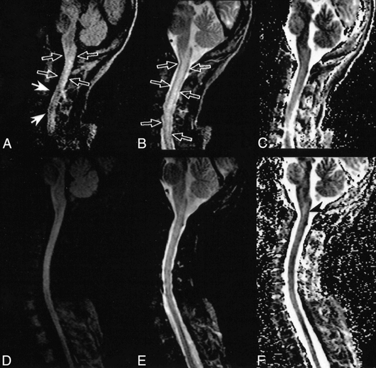
  • fig 3.
    • Download figure
    • Open in new tab
    • Download powerpoint
    fig 3.

    Only mild hyperintensity was seen on diffusion-weighted MR images of a patient with myelitis.

    A, Diffusion-weighted interleaved echo-planar image shows diffuse signal hyperintensity and swelling of the cervical spinal cord (open arrows). Note the signal cancellation due to ghosting artifacts (closed arrows) caused by bulk motion.

    B, T2-weighted interleaved echo-planar image shows diffuse signal hyperintensity and swelling of the cervical spinal cord (open arrows), consistent with myelitis.

    C, Region-of-interest measurements on corresponding apparent diffusion coefficient maps reveal moderately increased apparent diffusion coefficient values. Therefore, hyperintensity in diffusion-weighted MR imaging seems to be related to the so-called “T2-shine-through” effect that compensates for increased apparent diffusion coefficient.

    D, Diffusion-weighted interleaved echo-planar image obtained at follow-up 7 months later. No pathologic signal alteration is seen.

    E, T2-weighted interleaved echo-planar image obtained at follow-up 7 months later. The cord volume appears normal, and only faint intramedullary hyperintensities have remained.

    F, Apparent diffusion coefficient map shows a small area with elevated apparent diffusion coefficient at the level between the medulla oblongata and C1 (arrowhead), which may represent residual damage.

PreviousNext
Back to top

In this issue

American Journal of Neuroradiology
Vol. 21, Issue 3
1 Mar 2000
  • Table of Contents
  • Index by author
Advertisement
Print
Download PDF
Email Article

Thank you for your interest in spreading the word on American Journal of Neuroradiology.

NOTE: We only request your email address so that the person you are recommending the page to knows that you wanted them to see it, and that it is not junk mail. We do not capture any email address.

Enter multiple addresses on separate lines or separate them with commas.
Diffusion-Weighted MR Imaging of the Spinal Cord
(Your Name) has sent you a message from American Journal of Neuroradiology
(Your Name) thought you would like to see the American Journal of Neuroradiology web site.
CAPTCHA
This question is for testing whether or not you are a human visitor and to prevent automated spam submissions.
Cite this article
Roland Bammer, Franz Fazekas, Michael Augustin, Josef Simbrunner, Siegrid Strasser-Fuchs, Thomas Seifert, Rudolf Stollberger, Hans-Peter Hartung
Diffusion-Weighted MR Imaging of the Spinal Cord
American Journal of Neuroradiology Mar 2000, 21 (3) 587-591;

Citation Manager Formats

  • BibTeX
  • Bookends
  • EasyBib
  • EndNote (tagged)
  • EndNote 8 (xml)
  • Medlars
  • Mendeley
  • Papers
  • RefWorks Tagged
  • Ref Manager
  • RIS
  • Zotero
0 Responses
Respond to this article
Share
Bookmark this article
Diffusion-Weighted MR Imaging of the Spinal Cord
Roland Bammer, Franz Fazekas, Michael Augustin, Josef Simbrunner, Siegrid Strasser-Fuchs, Thomas Seifert, Rudolf Stollberger, Hans-Peter Hartung
American Journal of Neuroradiology Mar 2000, 21 (3) 587-591;
del.icio.us logo Twitter logo Facebook logo Mendeley logo
  • Tweet Widget
  • Facebook Like
  • Google Plus One

Jump to section

  • Article
    • Abstract
    • Description of the Technique
    • Discussion
    • Footnotes
    • References
  • Figures & Data
  • Info & Metrics
  • Responses
  • References
  • PDF

Related Articles

  • No related articles found.
  • PubMed
  • Google Scholar

Cited By...

  • Spinal cord infarction: a rare cause of paraplegia
  • Diffusion tensor imaging of the spinal cord and its clinical applications
  • Restricted Diffusion in Spinal Cord Infarction Demonstrated by Magnetic Resonance Line Scan Diffusion Imaging
  • Reduced Field-of-View Diffusion Imaging of the Human Spinal Cord: Comparison with Conventional Single-Shot Echo-Planar Imaging
  • Early diagnosis of spinal cord infarct using magnetic resonance diffusion imaging
  • Sensitivity-Encoded Diffusion Tensor MR Imaging of the Cervical Cord
  • Crossref
  • Google Scholar

This article has not yet been cited by articles in journals that are participating in Crossref Cited-by Linking.

More in this TOC Section

  • Management Outcomes For VO Spine Biopsy
  • Cone Beam CT Myelography
  • Post-Procedural Brachial Neuritis Features
Show more Spine Imaging and Spine Image-Guided Interventions

Similar Articles

Advertisement

Indexed Content

  • Current Issue
  • Accepted Manuscripts
  • Article Preview
  • Past Issues
  • Editorials
  • Editor's Choice
  • Fellows' Journal Club
  • Letters to the Editor
  • Video Articles

Cases

  • Case Collection
  • Archive - Case of the Week
  • Archive - Case of the Month
  • Archive - Classic Case

More from AJNR

  • Trainee Corner
  • Imaging Protocols
  • MRI Safety Corner
  • Book Reviews

Multimedia

  • AJNR Podcasts
  • AJNR Scantastics

Resources

  • Turnaround Time
  • Submit a Manuscript
  • Submit a Video Article
  • Submit an eLetter to the Editor/Response
  • Manuscript Submission Guidelines
  • Statistical Tips
  • Fast Publishing of Accepted Manuscripts
  • Graphical Abstract Preparation
  • Imaging Protocol Submission
  • Evidence-Based Medicine Level Guide
  • Publishing Checklists
  • Author Policies
  • Become a Reviewer/Academy of Reviewers
  • News and Updates

About Us

  • About AJNR
  • Editorial Board
  • Editorial Board Alumni
  • Alerts
  • Permissions
  • Not an AJNR Subscriber? Join Now
  • Advertise with Us
  • Librarian Resources
  • Feedback
  • Terms and Conditions
  • AJNR Editorial Board Alumni

American Society of Neuroradiology

  • Not an ASNR Member? Join Now

© 2025 by the American Society of Neuroradiology All rights, including for text and data mining, AI training, and similar technologies, are reserved.
Print ISSN: 0195-6108 Online ISSN: 1936-959X

Powered by HighWire