Article Figures & Data
Figures
FIG 1. Images from the case of a 6-year-old male patient.
A, Sagittal T1-weighted image from the right hemisphere shows perisylvian clustering of spike activity in the posterosuperior temporal gyrus or Wernicke's area.
B, Sagittal T1-weighted image from the left hemisphere shows perisylvian clustering of spike activity in the posterosuperior temporal gyrus or Wernicke's area.
C, Coronal T2-weighted image shows bilateral perisylvian clustering of spike activity in the posterosuperior temporal gyrus or Wernicke's area.
D, Six-second data epoch shows MEG wave forms from the left hemisphere above, right hemisphere below, and concurrent EEG in the middle. Numerous spikes are present bilaterally.
FIG 2. Image from the case of a 10-year-old female patient. Sagittal T1-weighted image shows unilateral clustering of spike activity in the left posterotemporal lobe, with spikes bordering the posterior aspect of the left sylvian fissure
Tables

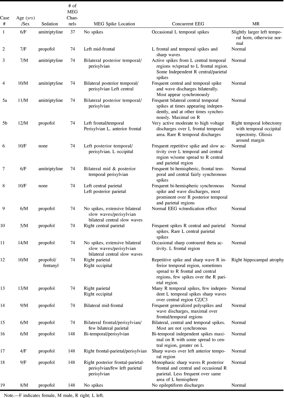
SUMMARY OF MEG, EEG and MR studies in 19 children with LKS and acquired epileptic aphasia


