Advertisement

Main menu

  • Home
  • Content
    • Current Issue
    • Accepted Manuscripts
    • Article Preview
    • Past Issue Archive
    • Video Articles
    • AJNR Case Collection
    • Case of the Week Archive
    • Case of the Month Archive
    • Classic Case Archive
  • Special Collections
    • Advancing NeuroMRI with High-Relaxivity Contrast Agents
    • AJNR Awards
    • Low-Field MRI
    • Alzheimer Disease
    • ASNR Foundation Special Collection
    • Photon-Counting CT
    • View All
  • Multimedia
    • AJNR Podcasts
    • AJNR SCANtastic
    • Trainee Corner
    • MRI Safety Corner
    • Imaging Protocols
  • For Authors
    • Submit a Manuscript
    • Submit a Video Article
    • Submit an eLetter to the Editor/Response
    • Manuscript Submission Guidelines
    • Statistical Tips
    • Fast Publishing of Accepted Manuscripts
    • Graphical Abstract Preparation
    • Imaging Protocol Submission
    • Author Policies
  • About Us
    • About AJNR
    • Editorial Board
    • Editorial Board Alumni
  • More
    • Become a Reviewer/Academy of Reviewers
    • Subscribers
    • Permissions
    • Alerts
    • Feedback
    • Advertisers
    • ASNR Home

User menu

  • Alerts
  • Log in

Search

  • Advanced search
American Journal of Neuroradiology

American Journal of Neuroradiology

ASHNR American Society of Functional Neuroradiology ASHNR American Society of Pediatric Neuroradiology ASSR
  • Alerts
  • Log in

Advanced Search

  • Home
  • Content
    • Current Issue
    • Accepted Manuscripts
    • Article Preview
    • Past Issue Archive
    • Video Articles
    • AJNR Case Collection
    • Case of the Week Archive
    • Case of the Month Archive
    • Classic Case Archive
  • Special Collections
    • Advancing NeuroMRI with High-Relaxivity Contrast Agents
    • AJNR Awards
    • Low-Field MRI
    • Alzheimer Disease
    • ASNR Foundation Special Collection
    • Photon-Counting CT
    • View All
  • Multimedia
    • AJNR Podcasts
    • AJNR SCANtastic
    • Trainee Corner
    • MRI Safety Corner
    • Imaging Protocols
  • For Authors
    • Submit a Manuscript
    • Submit a Video Article
    • Submit an eLetter to the Editor/Response
    • Manuscript Submission Guidelines
    • Statistical Tips
    • Fast Publishing of Accepted Manuscripts
    • Graphical Abstract Preparation
    • Imaging Protocol Submission
    • Author Policies
  • About Us
    • About AJNR
    • Editorial Board
    • Editorial Board Alumni
  • More
    • Become a Reviewer/Academy of Reviewers
    • Subscribers
    • Permissions
    • Alerts
    • Feedback
    • Advertisers
    • ASNR Home
  • Follow AJNR on Twitter
  • Visit AJNR on Facebook
  • Follow AJNR on Instagram
  • Join AJNR on LinkedIn
  • RSS Feeds

AJNR Awards, New Junior Editors, and more. Read the latest AJNR updates


Improved Turnaround Times | Median time to first decision: 12 days

Research ArticleNeurointervention

Angiographic Anatomy of the Laterocavernous Sinus

Philippe Gailloud, Diego San Millán Ruíz, Michel Muster, Kieran J. Murphy, Jean H. D. Fasel and Daniel A. Rüfenacht
American Journal of Neuroradiology November 2000, 21 (10) 1923-1929;
Philippe Gailloud
  • Find this author on Google Scholar
  • Find this author on PubMed
  • Search for this author on this site
Diego San Millán Ruíz
  • Find this author on Google Scholar
  • Find this author on PubMed
  • Search for this author on this site
Michel Muster
  • Find this author on Google Scholar
  • Find this author on PubMed
  • Search for this author on this site
Kieran J. Murphy
  • Find this author on Google Scholar
  • Find this author on PubMed
  • Search for this author on this site
Jean H. D. Fasel
  • Find this author on Google Scholar
  • Find this author on PubMed
  • Search for this author on this site
Daniel A. Rüfenacht
  • Find this author on Google Scholar
  • Find this author on PubMed
  • Search for this author on this site
  • Article
  • Figures & Data
  • Info & Metrics
  • Responses
  • References
  • PDF
Loading

Article Figures & Data

Figures

  • Tables
  • fig 1.
    • Download figure
    • Open in new tab
    • Download powerpoint
    fig 1.

    Schematic representation of the three basic drainage pathways of the SMCV according to San Millán Ruíz et al (4). Superior view of the anterior, middle, and posterior cranial fossa (ACF, MCF, and PCF, respectively). The SMCV may continue as a paracavernous sinus coursing laterally over the MCF (a), as an LCS enclosed within the lateral wall of the CS (b), or may terminate into the anterosuperior aspect of the CS (c). Venous outflow toward the pterygoid plexus via a skull base foramen is shown for the LCS. 1, superior ophthalmic vein; 2, CS; 3, inferior petrosal sinus; 4, sigmoid sinus; 5, transverse sinus; 6, SMCV

  • fig 2.
    • Download figure
    • Open in new tab
    • Download powerpoint
    fig 2.

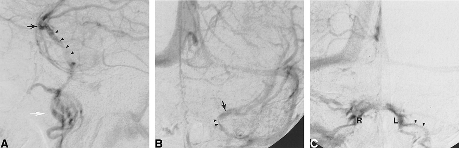
    DSA, venous phase, in a 59-year-old man examined for carotid bifurcation atheromatous disease. Intracranial vasculature is normal.

    A, Left ICA injection, lateral view. The left SMCV (black arrow) continues as an LCS (arrowheads), coursing posteriorly and inferiorly and projecting over the sellar region. In this case, note that the LCS drains toward the pterygoid plexus (white arrow) through foramina in the middle cranial fossa floor.

    B, Left ICA injection, AP view. The left SMCV (arrow) is opacified, as is a left LCS (arrowheads). There is no opacification of the left CS.

    C, Right ICA injection, AP view. Venous blood coming from the right cerebral hemisphere opacifies both the right CS (R) and left CS (L) through intercavernous connections. Note also opacification of the left inferior petrosal sinus (arrowheads), which was not visible on the left ICA injection.

  • fig 3.
    • Download figure
    • Open in new tab
    • Download powerpoint
    fig 3.

    Schematic representation of the laterosellar venous spaces, coronal view. The medial and lateral compartments of the CS and the LCS are shown in different shades of gray, as indicated at the bottom of the figure. The medial and lateral compartments of the CS are plexiform groups of veins lying on the medial and lateral aspects of the ICA, respectively. The LCS is a venous structure showing the morphologic characteristics of a dural sinus, located in between the outer (A) and inner (B) layers of the lateral wall of the CS. ICA indicates cavernous segment of the ICA; MCF, middle cranial fossa; P, pituitary gland; SB, sphenoid bone and sinus

  • fig 4.
    • Download figure
    • Open in new tab
    • Download powerpoint
    fig 4.

    DSA, venous phase, in a 52-year-old woman examined for subarachnoid hemorrhage.

    A, Right ICA injection, AP view. A duplicated SMCV (black arrows) runs medially toward the CS region to continue its course as an LCS (arrowheads), which terminates into the posterior aspect of the right CS via a small anastomotic channel (white arrow). The medial (M) and lateral (L) compartments of the CS and the right inferior petrosal sinus are well delineated. The left CS and inferior petrosal sinus are faintly opacified through intercavernous connections. The inner layer of the lateral wall of the CS is well appreciated as a curvilinear opacification defect located between the LCS and the lateral compartment of the CS.

    B, Right ICA injection, AP view, sequential subtraction technique. Simultaneous visualization of the arterial (light) and venous (dark) phases confirms that the small anastomotic channel connects the LCS to the lateral compartment of the CS, the latter abutting the lateral aspect of the cavernous segment of the left ICA.

  • fig 5.
    • Download figure
    • Open in new tab
    • Download powerpoint
    fig 5.

    DSA, venous phase, in a 46-year-old man examined for subarachnoid hemorrhage. Cerebral angiogram showed an anterior communicating artery aneurysm.

    A, Right internal carotid injection, AP view, sequential subtraction technique. The right SMCV (arrow) courses medially toward the CS region and continues posteriorly as an LCS (arrowheads). The LCS is lateral to the ICA, but not in its immediate proximity, and assumes a slitlike appearance. Since there is no connection between the LCS and the CS, the latter do not opacify and the LCS is consequently well appreciated. When connections allow for opacification of both the LCS and the CS, a slitlike LCS may be difficult to observe angiographically. Note the saccular aneurysm located at the right A1/A2 anterior communicating artery junction.

    B, Right internal carotid injection, lateral view. Owing to its thin configuration, the slitlike LCS (arrowheads) faintly opacifies on a lateral view. Note the drainage pathway toward the pterygoid plexus (arrow).

  • fig 6.
    • Download figure
    • Open in new tab
    • Download powerpoint
    fig 6.

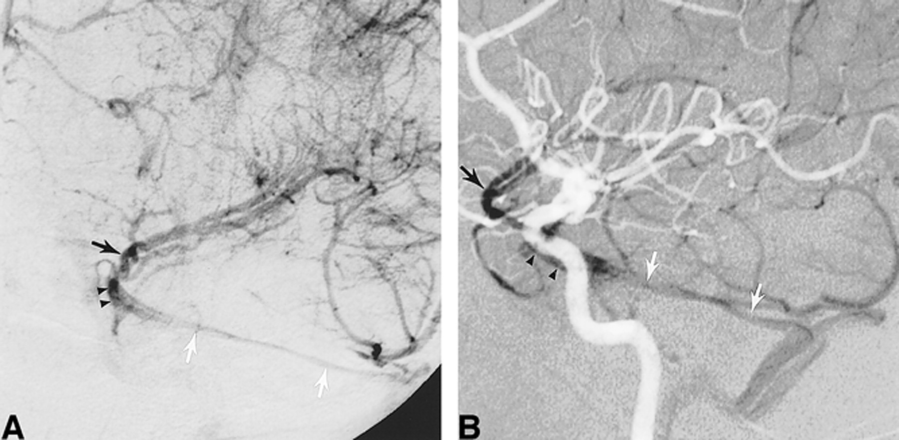
    DSA, venous phase, in a 38-year-old man examined for subarachnoid hemorrhage.

    A, Left ICA injection, left anterior oblique view. The left SMCV (black arrow) courses medially toward the CS region but continues posteriorly as an LCS (arrowheads) and then as an SPS (white arrows), ending in the left transverse sinus. There is no connection with the CS, which is not opacified.

    B, Left ICA injection, lateral view, sequential subtraction technique. The arterial (light) and venous (dark) phases are displayed simultaneously, enabling visualization of their topographic relationship. The LCS (arrowheads) is seen overlying the cavernous segment of the left ICA. Posteriorly, it continues as the left SPS (white arrows) coursing over the petrous ridge before reaching the left transverse sinus. Black arrow indicates the left SMCV.

  • fig 7.
    • Download figure
    • Open in new tab
    • Download powerpoint
    fig 7.

    DSA, venous phase, in a 37-year-old woman undergoing a presurgical epilepsy workup (Wada test).

    A, Right internal carotid injection, AP view. The right SMCV (arrow) drains toward the right transverse sinus via an LCS. A small anastomotic channel (arrowhead) joins the LCS to the posterior aspect of the CS at the skull base.

    B, Right internal carotid injection, lateral view. The simultaneous opacification of the LCS and the CS (medial and lateral compartments) makes it impossible to differentiate them on the lateral view.

    C, Left internal carotid injection, AP view. The left SMCV (arrow) drains toward the left pterygoid plexus (PP) via an LCS (arrowheads). There is an “en passant” connection between the LCS and the lateral compartment (L) of the CS.

  • fig 8.
    • Download figure
    • Open in new tab
    • Download powerpoint
    fig 8.

    DSA, venous phase, in a 68-year-old man examined for carotid bifurcation atheromatous disease.

    A, Right common carotid injection, AP view. The left SMCV (black arrow) drains into a right LCS (arrowheads). The right CS is opacified as well, although no significant connections are observed. Note the linear opacification defect of the inner layer of the CS lateral wall, separating the LCS from the lateral compartment of the CS. A second, round opacification defect is visible within the CS itself, corresponding to the cavernous segment of the right ICA (white arrow). This anatomic landmark enables one to discern the medial and lateral compartments of the CS.

    B, Magnified view of the right laterosellar venous spaces. This dynamic sequence (1 to 6) was acquired at the rate of one image per second. Note that the LCS appears early in the sequence while the medial and lateral compartments of the CS show homogeneous opacification after a delay of approximately 5 seconds. This is consistent with the findings of Bonneville et al (2), who observed, during dynamic CT of the laterosellar venous spaces, a delay of 5 seconds between the visualization of distinct laterosellar veins and a homogeneous opacification of the CS. Note on the last image of the sequence the particularly well delineated inner layer of the CS lateral wall.

Tables

  • Figures
  • TABLE 1:
    • View popup
    • Download powerpoint
    TABLE 1:

    Indication for cerebral angiography in the studied patient population

  • TABLE 2:
    • View popup
    • Download powerpoint
    TABLE 2:

    Drainage patterns of the paracavernous and laterocavernous sinuses

PreviousNext
Back to top

In this issue

American Journal of Neuroradiology
Vol. 21, Issue 10
1 Nov 2000
  • Table of Contents
  • Index by author
Advertisement
Print
Download PDF
Email Article

Thank you for your interest in spreading the word on American Journal of Neuroradiology.

NOTE: We only request your email address so that the person you are recommending the page to knows that you wanted them to see it, and that it is not junk mail. We do not capture any email address.

Enter multiple addresses on separate lines or separate them with commas.
Angiographic Anatomy of the Laterocavernous Sinus
(Your Name) has sent you a message from American Journal of Neuroradiology
(Your Name) thought you would like to see the American Journal of Neuroradiology web site.
CAPTCHA
This question is for testing whether or not you are a human visitor and to prevent automated spam submissions.
Cite this article
Philippe Gailloud, Diego San Millán Ruíz, Michel Muster, Kieran J. Murphy, Jean H. D. Fasel, Daniel A. Rüfenacht
Angiographic Anatomy of the Laterocavernous Sinus
American Journal of Neuroradiology Nov 2000, 21 (10) 1923-1929;

Citation Manager Formats

  • BibTeX
  • Bookends
  • EasyBib
  • EndNote (tagged)
  • EndNote 8 (xml)
  • Medlars
  • Mendeley
  • Papers
  • RefWorks Tagged
  • Ref Manager
  • RIS
  • Zotero
0 Responses
Respond to this article
Share
Bookmark this article
Angiographic Anatomy of the Laterocavernous Sinus
Philippe Gailloud, Diego San Millán Ruíz, Michel Muster, Kieran J. Murphy, Jean H. D. Fasel, Daniel A. Rüfenacht
American Journal of Neuroradiology Nov 2000, 21 (10) 1923-1929;
del.icio.us logo Twitter logo Facebook logo Mendeley logo
  • Tweet Widget
  • Facebook Like
  • Google Plus One

Jump to section

  • Article
    • Abstract
    • Methods
    • Results
    • Discussion
    • Conclusion
    • Footnotes
    • References
  • Figures & Data
  • Info & Metrics
  • Responses
  • References
  • PDF

Related Articles

  • No related articles found.
  • PubMed
  • Google Scholar

Cited By...

  • Non-aggressive, sinus-type greater sphenoid wing dural arteriovenous fistula with shunt point in the laterocavernous sinus mimicking a cavernous sinus dural arteriovenous fistula converted to aggressive, non-sinus-type
  • Cerebral venous anatomy: implications for the neurointerventionalist
  • Cerebral venous anatomy: implications for the neurointerventionalist
  • Republished: Embolization of a cavernous carotid fistula through the vein of Labbe: a new alternative transvenous access route
  • Embolization of a cavernous carotid fistula through the vein of Labbe: a new alternative transvenous access route
  • New Concept in Cavernous Sinus Dural Arteriovenous Fistula: Correlation With Presenting Symptom and Venous Drainage Patterns
  • The Sphenoparietal Sinus of Breschet: Does It Exist? An Anatomic Study
  • Crossref
  • Google Scholar

This article has not yet been cited by articles in journals that are participating in Crossref Cited-by Linking.

More in this TOC Section

  • Prophylactic Endovascular Treatments for CBS
  • Smoking & Aneurysm Outcomes: A Meta-Analysis
  • “Buddy-wire anchoring” technique for TVE
Show more NEUROINTERVENTION

Similar Articles

Advertisement

Indexed Content

  • Current Issue
  • Accepted Manuscripts
  • Article Preview
  • Past Issues
  • Editorials
  • Editor's Choice
  • Fellows' Journal Club
  • Letters to the Editor
  • Video Articles

Cases

  • Case Collection
  • Archive - Case of the Week
  • Archive - Case of the Month
  • Archive - Classic Case

More from AJNR

  • Trainee Corner
  • Imaging Protocols
  • MRI Safety Corner
  • Book Reviews

Multimedia

  • AJNR Podcasts
  • AJNR Scantastics

Resources

  • Turnaround Time
  • Submit a Manuscript
  • Submit a Video Article
  • Submit an eLetter to the Editor/Response
  • Manuscript Submission Guidelines
  • Statistical Tips
  • Fast Publishing of Accepted Manuscripts
  • Graphical Abstract Preparation
  • Imaging Protocol Submission
  • Evidence-Based Medicine Level Guide
  • Publishing Checklists
  • Author Policies
  • Become a Reviewer/Academy of Reviewers
  • News and Updates

About Us

  • About AJNR
  • Editorial Board
  • Editorial Board Alumni
  • Alerts
  • Permissions
  • Not an AJNR Subscriber? Join Now
  • Advertise with Us
  • Librarian Resources
  • Feedback
  • Terms and Conditions
  • AJNR Editorial Board Alumni

American Society of Neuroradiology

  • Not an ASNR Member? Join Now

© 2025 by the American Society of Neuroradiology All rights, including for text and data mining, AI training, and similar technologies, are reserved.
Print ISSN: 0195-6108 Online ISSN: 1936-959X

Powered by HighWire