
Article Figures & Data
Figures
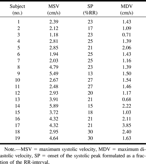
- fig 1.
Cervical syringomyelia. Axial slice, preoperative velocity imaging (patient 11).
Six images are obtained 30, 100, 150, 200, 300, and 400 ms after the R-wave. Craniocaudal velocities are represented in red and caudocranial velocites in blue. A pulsatile flow is observed as well in the cyst as in the PCSS.
- fig 2.
Normal kinetics of CSF at C2 and C6 levels (volunteer 15).
Caudal flow is represented by negative values. A systolic velocity peak is clearly defined, and it occurs in the first third of the RR cycle duration.
fig 3. Cystic and pericystic kinetics (patient 1).
A systolic peak is observed in the PCSS and in the cyst, but it occurs sooner in the cyst.
- fig 4.
Pre- and postoperative evolution (patient 13). Pre- and postoperative cyst morphology (A), pre- (B) and postoperative (C), velocity imaging, evolution of cyst (D), and of PCSS velocities (E) are shown. PCCS velocity increases in the postoperative course. In this case with a partial reduction of the cyst volume, cyst velocity distinctly decreases


Tables

In this issue


Jump to section
Related Articles
Cited By...
- MRI T2-Hyperintense Signal Structures in the Cervical Spinal Cord: Anterior Median Fissure versus Central Canal in Chiari and Control--An Exploratory Pilot Analysis
- Familial Adhesive Arachnoiditis Associated with Syringomyelia
- Cerebrospinal fluid flow imaging by using phase-contrast MR technique
- Characterization of CSF Hydrodynamics in the Presence and Absence of Tonsillar Ectopia by Means of Computational Flow Analysis
- Effect of Craniocervical Decompression on Peak CSF Velocities in Symptomatic Patients with Chiari I Malformation