
Article Figures & Data
Figures
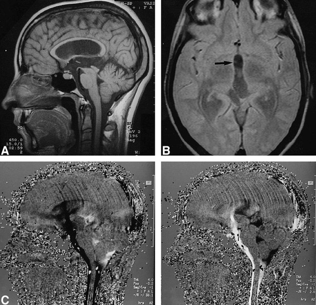
- fig 1.
Aqueductal stenosis due to presumed benign tectal glioma.
Sagittal midline contrast-enhanced T1-weighted image [(SE 450/15/2)(TR/TE/excitations)] shows nonenhancing bulbous enlargement of tectum (arrowhead), obliterating aqueduct and producing severe hydrocephalus. A small gap in floor of third ventricle in front of mamillary bodies can be identified (arrow).
B. Coronal (left) and sagittal (right) T2-weighted images (FSE 5000/90/2) show no flow-void across floor of third ventricle.
C. Proton-density-weighted axial image (SE 2200/20/1) at level of lateral ventricles shows marked hydrocephalus but no periventricular edema, indicating acute obstructive hydrocephalus.
D. Sagittal midline phase-contrast MR imaging [(2D-FISP 70/13/15°) (TR/TE/flip angle)]. Images represent a point in midsystole (left) and middiastole (right). Pulsatile CSF flow is seen within anterior of third ventricle and pontine cistern. CSF flow is rapid in anterior and slow in posterior of third ventricle, and absent through aqueduct.
E. Follow-up sagittal midline T1-weighted image (SE 450/15/2) performed after 4 years shows no changes in ventricular size and tectal mass.
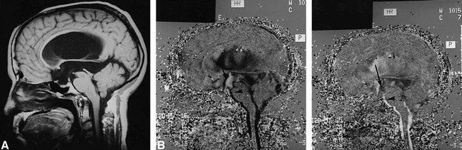
- fig 2.
Aqueductal stenosis.
A. Sagittal midline T1-weighted image (SE 450/15/2) reveals moderate ventricular dilatation with diffuse narrowing of aqueduct.
B. Proton-density-weighted axial image (SE 2200/20/1) at level of third ventricle shows abnormal flow-void at anterior recesses of third ventricle (arrow).
C. Sagittal midline phase-contrast MR imaging (2D-FISP 70/13/15°). A midsystolic image (left) shows caudal flow (black signal) at level of floor of third ventricle and prepontine cistern, while in middiastolic image (right) flow is reversed (white signal), indicating bidirectional flow through floor of third ventricle.
- fig 3.
Aqueductal stenosis.
A. Midline sagittal T1-weighted image (SE 550/14/2) in patient with moderate hydrocephalus shows distal aqueductal stenosis (arrow).
B. Proton-density axial image (SE 2200/20/1) at level of lateral ventricles shows moderate hydrocephalus but no periventricular edema, indicating acute obstructive hydrocephalus.
C–D. Sagittal midline phase-contrast MR imaging (2D-FISP 70/13/15°). Images represent a point in mid-systole (C) and middiastole (D). Craniocaudal pulsatile flow is seen through floor of third ventricle.
- fig 4.
Normal CSF flow cine-MR study. Sagittal midline phase-contrast MR imaging (2D-FISP 70/13/15°). Midsystolic image (left) shows caudal flow within foramen of Monro (arrow), aqueduct, and fourth ventricle. No CSF flow is seen in anterior of third ventricle. In middiastolic image (right), flow is reversed indicating bidirectional flow through ventricular system and subarachnoid spaces. By convention, caudal flow (systolic) is represented in black and cranial flow (diastolic) is represented in white
- fig 5.
Aqueductal stenosis without spontaneous ventriculostomy in patient with active obstructive hydrocephalus that required ventricular shunting.
A. Sagittal midline T1-weighted image (SE 450/15/2) reveals severe ventricular dilatation with distal narrowing of aqueduct.
B. Sagittal midline phase-contrast MR imaging (2D-FISP 70/13/15°). Images represent a point in midsystole (left) and middiastole (right). Craniocaudal pulsatile flow is not seen through aqueduct. Note asynchronic CSF flow between anteroinferior part of third ventricle (arrow) and prepontine subarachnoid spaces (arrowhead).


In this issue


Jump to section
Related Articles
Cited By...
- No citing articles found.