
Article Figures & Data
Figures
- fig 1.
14-year-old boy with pontine glioma. The T1-FSE sequence was performed before the T1-SE sequence.
A, Enhancing lesion cannot be identified on the T1-SE image (arrow).
B, Enhancement is seen within the pons on the T1-FSE image (arrow).
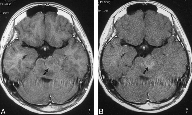
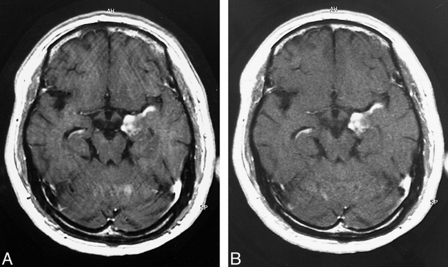
- fig 2.
60-year-old man with anaplastic ganglioglioma. The T1-FSE sequence was performed before the T1-SE sequence.
A, Peritumoral edema is present on the T1-SE image (arrow).
B, Peritumoral edema is inconspicuous on the T1-FSE image (arrow).
- fig 3.
45-year-old man with glioblastoma. The T1-FSE sequence was performed after the T1-SE sequence.
A and B, The T1-SE image (A) has more severe motion artifacts than does the T1-FSE image (B).
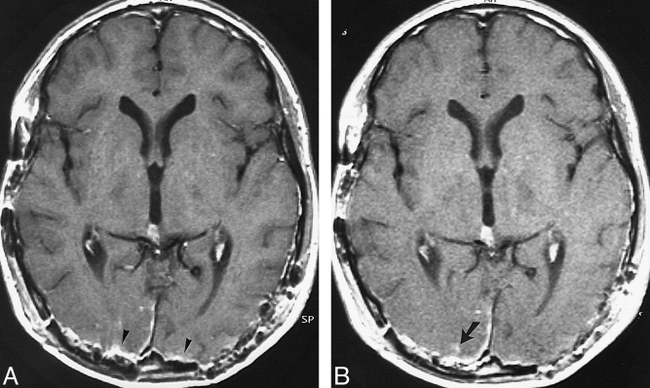
- fig 4.
16-year-old boy with germ cell tumor after surgery. The T1-FSE sequence was performed after the T1-SE sequence.
A, Metal artifacts (arrowheads) associated with surgery are present bilaterally in the occipital region on the T1-SE image.
B, Minimal intraparechymal enhancement, which was difficult to identify on the T1-SE image, is clearly depicted on the T1-FSE image (arrow).
- fig 5.
19-year-old man with multiple sclerosis. The T1-FSE sequence was performed before the T1-SE sequence.
A, On the T1-SE image, multiple sclerosis plaque is observed in right frontal white matter (arrow). No apparent enhancing lesion is visible.
B, The T1-FSE image shows hyperintense rim of multiple sclerosis plaque (arrow), which was not visible on the T1-SE image.


Tables
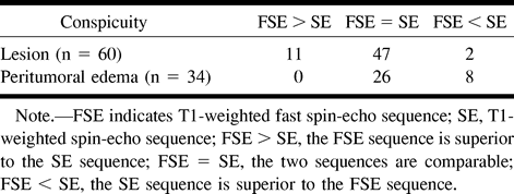
 TABLE 1:
Conspicuity of enhancing lesion and peritumoral edema in 60 patients with intracranial lesions
In this issue


Jump to section
Related Articles
Cited By...
- No citing articles found.