
Article Figures & Data
Figures
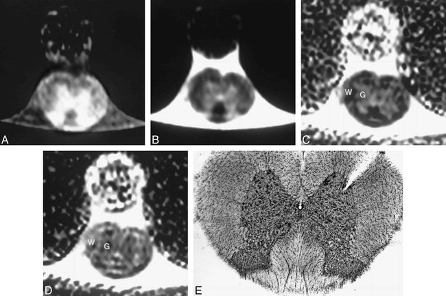
- fig 1.
A–E, Cross-sectional T1-weighted (800/22/6) (A) and T2-weighted (2000/80/4) (B) images of the spinal cord control specimen show expected hypointensity of white matter on both imaging sequences. Cross-sectional ADC images of this same animal in the read (C) and slice-select (D) axes show gray (G)/white (W) differentiation. A 2-mm dowel is seen ventrally. As expected, the gray matter is relatively isointense on both images but the white matter shows longitudinal anisotropy; ie, hypointense on the read axis (C) and hyperintense on the slice-select axis (D). Corresponding histopathologic section (E) is provided for images shown in A–D; the notch in the upper left of the specimen is used to mark the left side of the spinal cord before sectioning
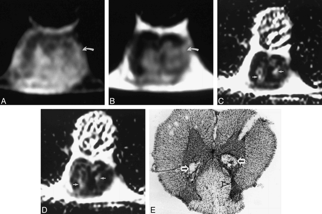
- fig 2.
A–E, Cross-sectional T1-weighted (800/22/6) (A) and T2-weighted (2000/80/4) (B) images of rat spinal cord 1 week after injection show abnormal heterogeneous signal in the gray matter, worse on the left, with associated loss of normal gray/white differentiation (curved arrow). Corresponding ADC maps in the read (C) and slice-select (D) axes show two areas of increased signal (arrows) in both axes, corresponding to the two cavities partially filled with cellular debris on the histopathologic specimen (arrows, E). The increased neuronal damage on the left is seen as a relative decrease in size of the dorsal gray matter on the left side of the spinal cord (arrowhead)
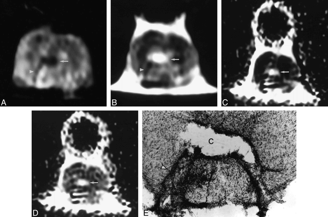
- fig 3.
A–E, Cross-sectional T1-weighted (800/22/6) (A) and T2-weighted (2000/80/4) (B) images of rat spinal cord 4 weeks after injection show a cystic lesion within the spinal cord (arrow), hypointense on the T1-weighted image and hyperintense on the T2-weighted image. A linear area of low signal intensity on both the T1- and T2-weighted images is seen within the dorsal gray matter (arrowhead), representing neuronal degeneration, gliosis, and scarring along the injection tract. Corresponding ADC maps in the read (C) and slice-select (D) axes show this cystic lesion (arrow) to be hyperintense in both axes. The cystic lesion seen on the MR images corresponds to the cyst (C) seen in the histopathologic specimen (E), and the glial scarring seen on the conventional MR images is marked with an arrowhead; note the decreased amount of debris within the cyst as compared with the cavities seen in figure 2E
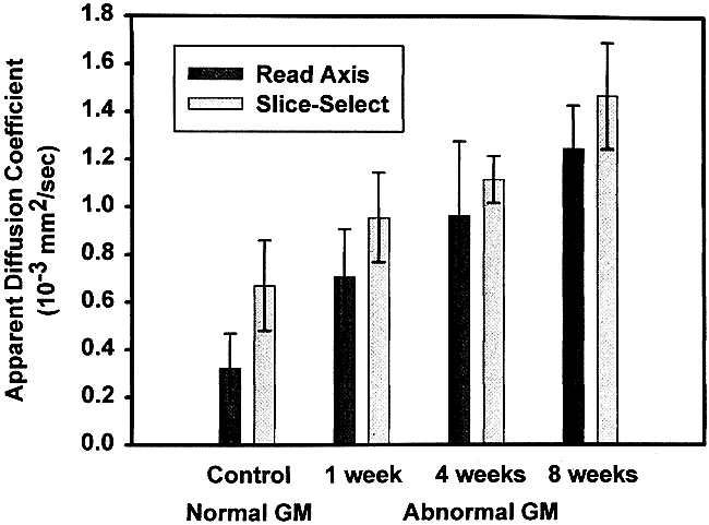
- fig 4.
Graph of the ADC values (10−3 mm2/s) in both the read and slice-select axes versus time. There is a progressive increase in ADC values (y-axis) of the lesions seen within the affected gray matter as the time (x-axis) since injection increased from 1 to 8 weeks


