
Article Figures & Data
Figures
- fig 1.
The hybrid 2D-CSI/1D-HSI sequence. A 25.6-millisecond 60-Hz CHESS sequence was followed by three 5.12-millisecond, dual-lobe, time-shifted 90+εi° (εi = 17°, 10°, and 5° to compensate for partial fat T1 recovery) OVS pulses. The VOI was selectively excited by a TR/TE = 1600/135 PRESS sequence with its 5.12-millisecond 90° pulse as well as fourth-order HSI encoding under a 3 mTM−1 gradient. This was followed 27.5 and 100 milliseconds later by a 5.12-millisecond 180° RF pulse under 1 mTM−1. The 2D 16 × 16 CSI sequence was performed during the TE along the x and y axes.
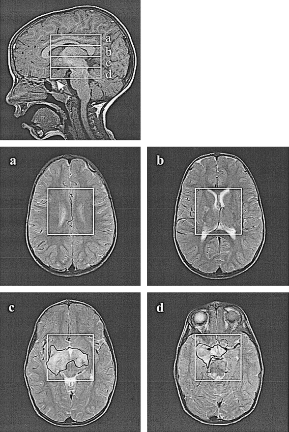
- fig 2.
Top, Sagittal image shows the placement of the 6 × 6 × 6-cm PRESS VOI, the four 1.5-cm-thick HSI slices, a–d, and the chiasmal glioma (arrow) in a 3-year-old boy.
a–d, Axial T2-weighted images (FOV = 22 cm) of the corresponding HSI slices, superimposed with the axial projection of the 6 × 6-cm VOIs. The FASI regions were marked with a solid black line by a neuroradiologist. The dotted circle in d indicates the location of the glioma.
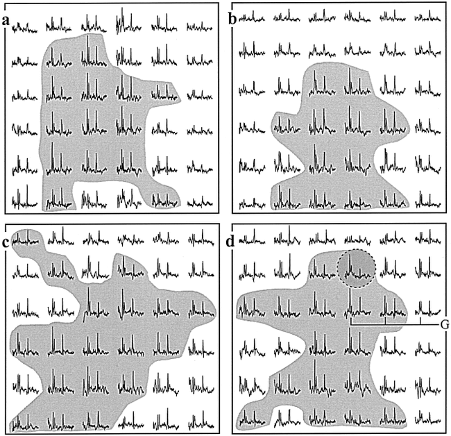
- fig 3.
a–d, Real part of the proton spectra from the PRESS boxes on the corresponding images in figure 2. The tumor spectrum in d is highlighted with a dashed shaded circle, and regions of elevated 2>Cho:Cr>1.3 are shaded. All spectra display the 4.0 to 0.5 ppm chemical shift range and are plotted on the same vertical scale, and each represents signal from a 1.5-cm3 voxel
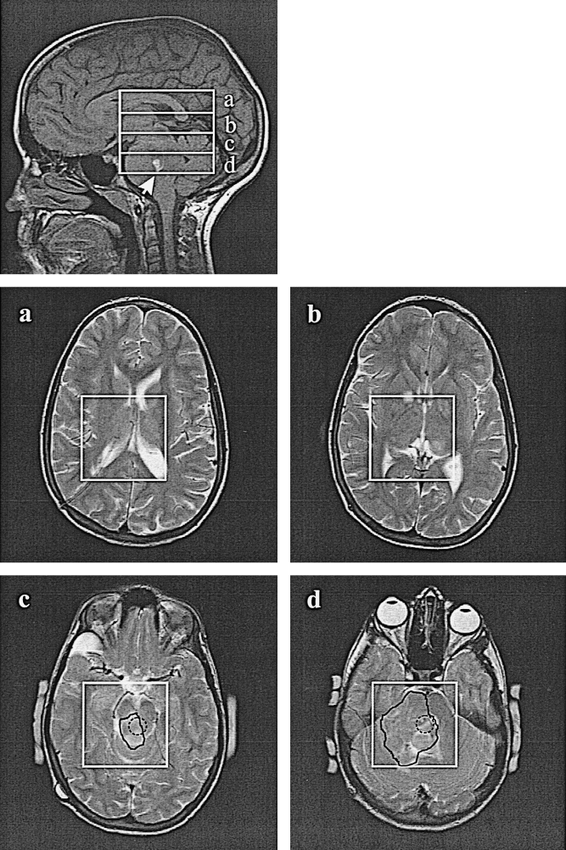
- fig 4.
Top, Sagittal image superimposed with the 6 × 6 × 6-cm PRESS VOI, the four 1.5-cm-thick HSI slices, a–d, and the brain stem glioma in an 11-year-old girl (arrow).
a–d, PRESS VOIs and FASI regions are marked with a solid black line on the corresponding axial T2-weighted images (FOV = 22 cm) from slices a–d. The dotted circles on axial images c and d indicate the location of the glioma.
- fig 5.
a–d, Real part of the proton spectra from the VOIs on the corresponding images in figure 4. The spectra from the tumor in slices c and d in the dashed circle and regions of elevated 2>Cho:Cr>1.3 are shaded. All spectra display the 4.0 to 0.5 ppm chemical shift range and are plotted on the same vertical scale, and each represents the signal from a 1.5-cm3 voxel
- fig 6.
Top row, Axial images show the location of the PRESS boxes and 1 × 1-cm CSI grid from the brain of a healthy male 7-month-old control subject, corresponding to the patient in figures 2c and d and 3c and d.
Bottom row, Real part of the 8 × 8 proton spectra matrices from the corresponding PRESS boxes. Spectra display the 4.0 to 0.5 ppm chemical shift range from each 1-cm3 voxel and are on a common intensity grid.
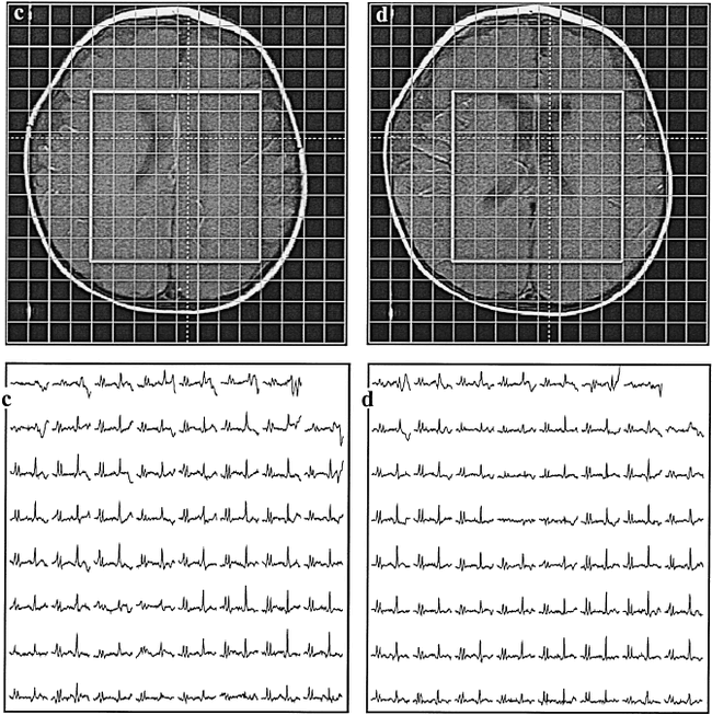
- fig 7.
Top, Axial images show the location of the PRESS boxes and 1 × 1-cm CSI grid from the brain of a healthy male 11-year-old control subject, corresponding to the patient in figures 4c and d and 5c and d.
Bottom, Real part of the 8 × 8 proton spectra matrices from the corresponding PRESS boxes. Spectra display the 4.0 to 0.5 ppm chemical shift range from each 1-cm3 voxel and are on a common intensity grid.


