Advertisement
  • Main menu
  • User menu
  • Search
  • English ▼
    • English
    • Afrikaans
    • Albanian
    • Amharic
    • Arabic
    • Armenian
    • Azerbaijani
    • Basque
    • Belarusian
    • Bengali
    • Bosnian
    • Bulgarian
    • Catalan
    • Cebuano
    • Chichewa
    • Chinese (Simplified)
    • Chinese (Traditional)
    • Corsican
    • Croatian
    • Czech
    • Danish
    • Dutch
    • Esperanto
    • Estonian
    • Filipino
    • Finnish
    • French
    • Frisian
    • Galician
    • Georgian
    • German
    • Greek
    • Gujarati
    • Haitian Creole
    • Hausa
    • Hawaiian
    • Hebrew
    • Hindi
    • Hmong
    • Hungarian
    • Icelandic
    • Igbo
    • Indonesian
    • Irish
    • Italian
    • Japanese
    • Javanese
    • Kannada
    • Kazakh
    • Khmer
    • Korean
    • Kurdish (Kurmanji)
    • Kyrgyz
    • Lao
    • Latin
    • Latvian
    • Lithuanian
    • Luxembourgish
    • Macedonian
    • Malagasy
    • Malay
    • Malayalam
    • Maltese
    • Maori
    • Marathi
    • Mongolian
    • Myanmar (Burmese)
    • Nepali
    • Norwegian
    • Pashto
    • Persian
    • Polish
    • Portuguese
    • Punjabi
    • Romanian
    • Russian
    • Samoan
    • Scottish Gaelic
    • Serbian
    • Sesotho
    • Shona
    • Sindhi
    • Sinhala
    • Slovak
    • Slovenian
    • Somali
    • Spanish
    • Sudanese
    • Swahili
    • Swedish
    • Tajik
    • Tamil
    • Telugu
    • Thai
    • Turkish
    • Ukrainian
    • Urdu
    • Uzbek
    • Vietnamese
    • Welsh
    • Xhosa
    • Yiddish
    • Yoruba
    • Zulu

Main menu

  • Home
  • Content
    • Current Issue
    • Accepted Manuscripts
    • Article Preview
    • Past Issue Archive
    • Video Articles
    • AJNR Case Collection
    • Case of the Week Archive
    • Case of the Month Archive
    • Classic Case Archive
  • Special Collections
    • Advancing NeuroMRI with High-Relaxivity Contrast Agents
    • AJNR Awards
    • Low-Field MRI
    • Alzheimer Disease
    • ASNR Foundation Special Collection
    • Photon-Counting CT
    • View All
  • Multimedia
    • AJNR Podcasts
    • AJNR SCANtastic
    • Trainee Corner
    • MRI Safety Corner
    • Imaging Protocols
  • For Authors
    • Submit a Manuscript
    • Submit a Video Article
    • Submit an eLetter to the Editor/Response
    • Manuscript Submission Guidelines
    • Statistical Tips
    • Fast Publishing of Accepted Manuscripts
    • Graphical Abstract Preparation
    • Imaging Protocol Submission
    • Author Policies
  • About Us
    • About AJNR
    • Editorial Board
    • Editorial Board Alumni
  • More
    • Become a Reviewer/Academy of Reviewers
    • Subscribers
    • Permissions
    • Alerts
    • Feedback
    • Advertisers
    • ASNR Home

User menu

  • Alerts
  • Log in

Search

  • Advanced search
American Journal of Neuroradiology

American Journal of Neuroradiology

ASHNR American Society of Functional Neuroradiology ASHNR American Society of Pediatric Neuroradiology ASSR
  • Alerts
  • Log in

Advanced Search

English ▼
  • English
  • Afrikaans
  • Albanian
  • Amharic
  • Arabic
  • Armenian
  • Azerbaijani
  • Basque
  • Belarusian
  • Bengali
  • Bosnian
  • Bulgarian
  • Catalan
  • Cebuano
  • Chichewa
  • Chinese (Simplified)
  • Chinese (Traditional)
  • Corsican
  • Croatian
  • Czech
  • Danish
  • Dutch
  • Esperanto
  • Estonian
  • Filipino
  • Finnish
  • French
  • Frisian
  • Galician
  • Georgian
  • German
  • Greek
  • Gujarati
  • Haitian Creole
  • Hausa
  • Hawaiian
  • Hebrew
  • Hindi
  • Hmong
  • Hungarian
  • Icelandic
  • Igbo
  • Indonesian
  • Irish
  • Italian
  • Japanese
  • Javanese
  • Kannada
  • Kazakh
  • Khmer
  • Korean
  • Kurdish (Kurmanji)
  • Kyrgyz
  • Lao
  • Latin
  • Latvian
  • Lithuanian
  • Luxembourgish
  • Macedonian
  • Malagasy
  • Malay
  • Malayalam
  • Maltese
  • Maori
  • Marathi
  • Mongolian
  • Myanmar (Burmese)
  • Nepali
  • Norwegian
  • Pashto
  • Persian
  • Polish
  • Portuguese
  • Punjabi
  • Romanian
  • Russian
  • Samoan
  • Scottish Gaelic
  • Serbian
  • Sesotho
  • Shona
  • Sindhi
  • Sinhala
  • Slovak
  • Slovenian
  • Somali
  • Spanish
  • Sudanese
  • Swahili
  • Swedish
  • Tajik
  • Tamil
  • Telugu
  • Thai
  • Turkish
  • Ukrainian
  • Urdu
  • Uzbek
  • Vietnamese
  • Welsh
  • Xhosa
  • Yiddish
  • Yoruba
  • Zulu
  • Home
  • Content
    • Current Issue
    • Accepted Manuscripts
    • Article Preview
    • Past Issue Archive
    • Video Articles
    • AJNR Case Collection
    • Case of the Week Archive
    • Case of the Month Archive
    • Classic Case Archive
  • Special Collections
    • Advancing NeuroMRI with High-Relaxivity Contrast Agents
    • AJNR Awards
    • Low-Field MRI
    • Alzheimer Disease
    • ASNR Foundation Special Collection
    • Photon-Counting CT
    • View All
  • Multimedia
    • AJNR Podcasts
    • AJNR SCANtastic
    • Trainee Corner
    • MRI Safety Corner
    • Imaging Protocols
  • For Authors
    • Submit a Manuscript
    • Submit a Video Article
    • Submit an eLetter to the Editor/Response
    • Manuscript Submission Guidelines
    • Statistical Tips
    • Fast Publishing of Accepted Manuscripts
    • Graphical Abstract Preparation
    • Imaging Protocol Submission
    • Author Policies
  • About Us
    • About AJNR
    • Editorial Board
    • Editorial Board Alumni
  • More
    • Become a Reviewer/Academy of Reviewers
    • Subscribers
    • Permissions
    • Alerts
    • Feedback
    • Advertisers
    • ASNR Home
  • Follow AJNR on Twitter
  • Visit AJNR on Facebook
  • Follow AJNR on Instagram
  • Join AJNR on LinkedIn
  • RSS Feeds

AJNR Awards, New Junior Editors, and more. Read the latest AJNR updates


Improved Turnaround Times | Median time to first decision: 12 days

Research ArticleNeurointervention

Definition of the Ostium (Neck) of an Aneurysm Revealed by Intravascular Sonography: An Experimental Study in Canines

Aquilla S. Turk, Charles M. Strother, Daniel I. Crouthamel and James A. Zagzebski
American Journal of Neuroradiology July 1999, 20 (7) 1301-1308;
Aquilla S. Turk
  • Find this author on Google Scholar
  • Find this author on PubMed
  • Search for this author on this site
Charles M. Strother
  • Find this author on Google Scholar
  • Find this author on PubMed
  • Search for this author on this site
Daniel I. Crouthamel
  • Find this author on Google Scholar
  • Find this author on PubMed
  • Search for this author on this site
James A. Zagzebski
  • Find this author on Google Scholar
  • Find this author on PubMed
  • Search for this author on this site
  • Article
  • Figures & Data
  • Info & Metrics
  • Responses
  • References
  • PDF
Loading

Article Figures & Data

Figures

  • fig 1.
    • Download figure
    • Open in new tab
    • Download powerpoint
    fig 1.

    Digital subtraction angiogram of lateral (LA) and bifurcation (BA) aneurysms in a canine model. This image was obtained with a portable C-arm device
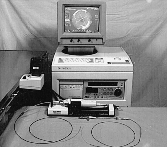
  • fig 2.
    • Download figure
    • Open in new tab
    • Download powerpoint
    fig 2.

    Intravascular sonographic machine with automated pullback servo and catheters
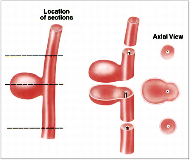
  • fig 3.
    • Download figure
    • Open in new tab
    • Download powerpoint
    fig 3.

    Left, Diagram of a lateral aneurysm and parent artery (dashed lines indicate locations in which intravascular sonographic measurements were made).

    Center, Line drawing of sections through the parent artery and the aneurysm in the plane of intravascular sonography.

    Right, Line drawing of corresponding intravascular sonograms.

  • fig 4.
    • Download figure
    • Open in new tab
    • Download powerpoint
    fig 4.

    Left, Diagram of a bifurcation aneurysm, parent artery, and adjacent branches (dashed lines indicate locations at which intravascular sonographic measurements were made).

    Center, Line drawings of sections through the parent artery, the aneurysm, and adjacent branches in the plane of intravascular sonography.

    Right, Line drawing of corresponding intravascular sonograms.

  • fig 5.
    • Download figure
    • Open in new tab
    • Download powerpoint
    fig 5.

    A, Angiogram of experimental aneurysm. The projection is optimized to allow best possible visualization of the lateral aneurysm's neck.

    B, Intravascular sonogram of the parent artery that gives rise to the lateral aneurysm.

    C, Intravascular sonogram through the lateral aneurysmal ostium. This image illustrates the ability to depict the width of the ostium (arrowheads indicate the point at which the lateral aneurysm, LA, joins the parent artery, PA; dashed line denotes the width of the ostium). In the intravascular sonograms, the distance between white marker dots is 1 mm and the intravascular sonographic catheter is defined by an asterisk.

  • fig 6.
    • Download figure
    • Open in new tab
    • Download powerpoint
    fig 6.

    A, Angiogram of a lateral and a bifurcation aneurysm after treatment with GDCs. Coils are purposely positioned so as to protrude into the parent artery (arrowheads). A filling defect (arrow) along the wall of the parent artery of the lateral aneurysm represents thrombus.

    B, Intravascular sonogram at the level of the filling defect on a DSA clearly shows the thrombus (arrowhead) as hyperechoic signal within the lumen.

    C, Intravascular sonogram at the upper extent of the aneurysm's ostium shows coils filling the ostium (dashed line indicates limits of the aneurysmal ostium; X‘s identify the spaces between coils).

    D, Intravascular sonogram at the lower extent of the aneurysm's ostium shows a portion of the outflow tract that was not occluded (X) (dashed line defines the limits of the aneurysmal ostium). In the intravascular sonograms the distance between the white marker dots is 2 mm, and the intravascular sonographic catheter is defined by an asterisk.

  • fig 7.
    • Download figure
    • Open in new tab
    • Download powerpoint
    fig 7.

    A, Angiogram of experimental aneurysms. The projection is optimized to allow visualization of the bifurcation aneurysm's neck.

    B, Intravascular sonogram in the dome of the bifurcation aneurysm (arrowheads define the wall of the aneurysm).

    C, Intravascular sonogram at a location just on the aneurysm side of the ostium (Os). The adjacent right and left carotid artery branches (RCa and LCa, respectively) are seen on either side of the aneurysm's wall as hypoechoic regions (arrowheads define the wall of the aneurysm).

    D, Intravascular sonogram at the isthmus between the ostium (Os) and adjacent bifurcation branches (dashed lines indicate position at which ostium opens into the bifurcation of the left and right carotid arteries, LCa and RCa, respectively). In the intravascular sonograms the distance between white marker dots is 2 mm, and the intravascular sonographic catheter is defined by an asterisk.

  • fig 8.
    • Download figure
    • Open in new tab
    • Download powerpoint
    fig 8.

    A–D, 2D reformations of a bifurcation aneurysm in the coronal (A) and sagittal (B) planes with corresponding axial image at the ostium (C) and a DSA (D) (dashed lines indicate the limit of the ostium; BA, bifurcation aneurysm; PA, parent artery; RCa, right carotid artery; LCa, left carotid artery; Os, ostium; asterisk denotes the intravascular sonographic catheter)

  • fig 9.
    • Download figure
    • Open in new tab
    • Download powerpoint
    fig 9.

    A, 2D reformation of a lateral aneurysm in the coronal plane. The artifact in this image is caused by up-and-down motion of the intravascular sonographic catheter during the cardiac cycle (asterisk denotes the intravascular sonographic catheter; LA, lateral aneurysm; PA, parent artery; dashed line identifies the limit of the aneurysm ostia).

    B, Corresponding DSA.

  • fig 10.
    • Download figure
    • Open in new tab
    • Download powerpoint
    fig 10.

    Regression plots of inter- and intraobserver correlation coefficients (P < .0001 in each instance)

PreviousNext
Back to top

In this issue

American Journal of Neuroradiology
Vol. 20, Issue 7
1 Jul 1999
  • Table of Contents
  • Index by author
Advertisement
Print
Download PDF
Email Article
Cite this article
0 Responses
Respond to this article
Share
Bookmark this article
Definition of the Ostium (Neck) of an Aneurysm Revealed by Intravascular Sonography: An Experimental Study in Canines
Aquilla S. Turk, Charles M. Strother, Daniel I. Crouthamel, James A. Zagzebski
American Journal of Neuroradiology Jul 1999, 20 (7) 1301-1308;
del.icio.us logo Twitter logo Facebook logo Mendeley logo
  • Tweet Widget

Jump to section

  • Article
    • Abstract
    • Methods
    • Results
    • Discussion
    • Conclusion
    • Acknowledgments
    • Footnotes
    • References
  • Figures & Data
  • Info & Metrics
  • Responses
  • References
  • PDF

Related Articles

  • No related articles found.
  • PubMed
  • Google Scholar

Cited By...

  • Technology developments in endovascular treatment of intracranial aneurysms
  • INTERVENTIONAL NEURORADIOLOGY
  • Crossref
  • Google Scholar

This article has not yet been cited by articles in journals that are participating in Crossref Cited-by Linking.

More in this TOC Section

  • Prophylactic Endovascular Treatments for CBS
  • Impact of Stenosis Degree on Outcomes of Stenting
  • Connectivity for remote neurovascular procedures
Show more Neurointervention

Similar Articles

Advertisement

Indexed Content

  • Current Issue
  • Accepted Manuscripts
  • Article Preview
  • Past Issues
  • Editorials
  • Editor's Choice
  • Fellows' Journal Club
  • Letters to the Editor
  • Video Articles

Cases

  • Case Collection
  • Archive - Case of the Week
  • Archive - Case of the Month
  • Archive - Classic Case

More from AJNR

  • Trainee Corner
  • Imaging Protocols
  • MRI Safety Corner
  • Book Reviews

Multimedia

  • AJNR Podcasts
  • AJNR Scantastics

Resources

  • Turnaround Time
  • Submit a Manuscript
  • Submit a Video Article
  • Submit an eLetter to the Editor/Response
  • Manuscript Submission Guidelines
  • Statistical Tips
  • Fast Publishing of Accepted Manuscripts
  • Graphical Abstract Preparation
  • Imaging Protocol Submission
  • Evidence-Based Medicine Level Guide
  • Publishing Checklists
  • Author Policies
  • Become a Reviewer/Academy of Reviewers
  • News and Updates

About Us

  • About AJNR
  • Editorial Board
  • Editorial Board Alumni
  • Alerts
  • Permissions
  • Not an AJNR Subscriber? Join Now
  • Advertise with Us
  • Librarian Resources
  • Feedback
  • Terms and Conditions
  • AJNR Editorial Board Alumni

American Society of Neuroradiology

  • Not an ASNR Member? Join Now

© 2025 by the American Society of Neuroradiology All rights, including for text and data mining, AI training, and similar technologies, are reserved.
Print ISSN: 0195-6108 Online ISSN: 1936-959X

Powered by HighWire
Email this Article

Thank you for your interest in spreading the word on American Journal of Neuroradiology.

NOTE: We only request your email address so that the person you are recommending the page to knows that you wanted them to see it, and that it is not junk mail. We do not capture any email address.

Enter multiple addresses on separate lines or separate them with commas.
Definition of the Ostium (Neck) of an Aneurysm Revealed by Intravascular Sonography: An Experimental Study in Canines
(Your Name) has sent you a message from American Journal of Neuroradiology
(Your Name) thought you would like to see the American Journal of Neuroradiology web site.
CAPTCHA
This question is for testing whether or not you are a human visitor and to prevent automated spam submissions.
Cite this article
Aquilla S. Turk, Charles M. Strother, Daniel I. Crouthamel, James A. Zagzebski
Definition of the Ostium (Neck) of an Aneurysm Revealed by Intravascular Sonography: An Experimental Study in Canines
American Journal of Neuroradiology Jul 1999, 20 (7) 1301-1308;

Citation Manager Formats

  • BibTeX
  • Bookends
  • EasyBib
  • EndNote (tagged)
  • EndNote 8 (xml)
  • Medlars
  • Mendeley
  • Papers
  • RefWorks Tagged
  • Ref Manager
  • RIS
  • Zotero

We use cookies on this site to enhance your user experience. By clicking any link on this page you are giving your consent for us to set cookies.