Advertisement

Main menu

  • Home
  • Content
    • Current Issue
    • Accepted Manuscripts
    • Article Preview
    • Past Issue Archive
    • Video Articles
    • AJNR Case Collection
    • Case of the Week Archive
    • Case of the Month Archive
    • Classic Case Archive
  • Special Collections
    • Advancing NeuroMRI with High-Relaxivity Contrast Agents
    • AJNR Awards
    • Low-Field MRI
    • Alzheimer Disease
    • ASNR Foundation Special Collection
    • Photon-Counting CT
    • View All
  • Multimedia
    • AJNR Podcasts
    • AJNR SCANtastic
    • Trainee Corner
    • MRI Safety Corner
    • Imaging Protocols
  • For Authors
    • Submit a Manuscript
    • Submit a Video Article
    • Submit an eLetter to the Editor/Response
    • Manuscript Submission Guidelines
    • Statistical Tips
    • Fast Publishing of Accepted Manuscripts
    • Graphical Abstract Preparation
    • Imaging Protocol Submission
    • Author Policies
  • About Us
    • About AJNR
    • Editorial Board
    • Editorial Board Alumni
  • More
    • Become a Reviewer/Academy of Reviewers
    • Subscribers
    • Permissions
    • Alerts
    • Feedback
    • Advertisers
    • ASNR Home

User menu

  • Alerts
  • Log in

Search

  • Advanced search
American Journal of Neuroradiology

American Journal of Neuroradiology

ASHNR American Society of Functional Neuroradiology ASHNR American Society of Pediatric Neuroradiology ASSR
  • Alerts
  • Log in

Advanced Search

  • Home
  • Content
    • Current Issue
    • Accepted Manuscripts
    • Article Preview
    • Past Issue Archive
    • Video Articles
    • AJNR Case Collection
    • Case of the Week Archive
    • Case of the Month Archive
    • Classic Case Archive
  • Special Collections
    • Advancing NeuroMRI with High-Relaxivity Contrast Agents
    • AJNR Awards
    • Low-Field MRI
    • Alzheimer Disease
    • ASNR Foundation Special Collection
    • Photon-Counting CT
    • View All
  • Multimedia
    • AJNR Podcasts
    • AJNR SCANtastic
    • Trainee Corner
    • MRI Safety Corner
    • Imaging Protocols
  • For Authors
    • Submit a Manuscript
    • Submit a Video Article
    • Submit an eLetter to the Editor/Response
    • Manuscript Submission Guidelines
    • Statistical Tips
    • Fast Publishing of Accepted Manuscripts
    • Graphical Abstract Preparation
    • Imaging Protocol Submission
    • Author Policies
  • About Us
    • About AJNR
    • Editorial Board
    • Editorial Board Alumni
  • More
    • Become a Reviewer/Academy of Reviewers
    • Subscribers
    • Permissions
    • Alerts
    • Feedback
    • Advertisers
    • ASNR Home
  • Follow AJNR on Twitter
  • Visit AJNR on Facebook
  • Follow AJNR on Instagram
  • Join AJNR on LinkedIn
  • RSS Feeds

AJNR Awards, New Junior Editors, and more. Read the latest AJNR updates


Improved Turnaround Times | Median time to first decision: 12 days

Research ArticleBrain

Loss of Interhemispheric Connectivity in Patients with Lacunar Infarction Reflected by Diffusion-Weighted MR Imaging and Single-Photon Emission CT

Makiko Ishihara, Shin-ichiro Kumita, Hiromitsu Hayashi and Tatsuo Kumazaki
American Journal of Neuroradiology June 1999, 20 (6) 991-998;
Makiko Ishihara
  • Find this author on Google Scholar
  • Find this author on PubMed
  • Search for this author on this site
Shin-ichiro Kumita
  • Find this author on Google Scholar
  • Find this author on PubMed
  • Search for this author on this site
Hiromitsu Hayashi
  • Find this author on Google Scholar
  • Find this author on PubMed
  • Search for this author on this site
Tatsuo Kumazaki
  • Find this author on Google Scholar
  • Find this author on PubMed
  • Search for this author on this site
  • Article
  • Figures & Data
  • Info & Metrics
  • Responses
  • References
  • PDF
Loading

Article Figures & Data

Figures

  • Tables
  • fig 1.
    • Download figure
    • Open in new tab
    • Download powerpoint
    fig 1.

    A representative level of calculated ADC maps. ROIs, placed on the anterior and posterior corpus callosum and optic radiation, are 100 to 200 pixels each and the same in contour between the two maps.

    A, ADC

    B, ADC

    C, A scheme for regional data extraction from these maps.

  • fig 2.
    • Download figure
    • Open in new tab
    • Download powerpoint
    fig 2.

    A–D, A representative series of binary SPECT scans showing masking levels of 60% (A), 65% (B), and 70% (C) of the maximal counts and the original SPECT scan (D). As the masking level increased, the number of masked pixels of relatively low counts also increased

  • fig 3.
    • Download figure
    • Open in new tab
    • Download powerpoint
    fig 3.

    An explanation of how the ACV, determined as the ratio of the number of pixels above the 80% masking level to the number above the 70% level, is validated. In this setting, the ACVs derived from the eight cortical areas should be homogeneous, because the actual tracer accumulation in the cortical area is uniform regardless of the regions.

    A, Phantom MR image.

    B, Phantom 123I-IMP SPECT scan.
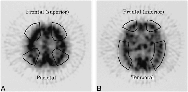
  • fig 4.
    • Download figure
    • Open in new tab
    • Download powerpoint
    fig 4.

    A and B, A representation of regional data extraction from one of the original SPECT scans. ROIs are specified at the superior frontal lobe and the parietal lobe (A), and the inferior frontal lobe and the temporal lobe (B), so as not to include the primary sensorimotor area, mesial temporal lobe, or insula. The ROIs are superimposed on ACV maps

  • fig 5.
    • Download figure
    • Open in new tab
    • Download powerpoint
    fig 5.

    Correlations between the regional ACV and the MMSE scores. Individual ACVs in the superior and inferior frontal regions were well correlated with the MMSE score (superior frontal: r = .89, P < .05; inferior frontal, r = .71, P < .05).

    Left upper, superior frontal.

    Right upper, inferior frontal.

    Left lower, parietal.

    Right lower, temporal.

  • fig 6.
    • Download figure
    • Open in new tab
    • Download powerpoint
    fig 6.

    Correlations between the regional AR of the corpus callosum and the ACV of the frontal areas. The AR in the anterior corpus callosum is strongly correlated with the frontal ACVs (superior frontal: r = .86, P < .0001; inferior frontal: r = .79, P < .0005). However, there are insignificant trends toward correlation between the AR in the anterior corpus callosum and the frontal ACVs within the patient group.

    Left upper, anterior corpus callosum versus superior frontal region.

    Right upper, anterior corpus callosum versus inferior frontal region.

    Left lower, posterior corpus callosum versus superior frontal region.

    Right lower, posterior corpus callosum versus inferior frontal region.

Tables

  • Figures
  • Table 1:
    • View popup
    • Download powerpoint
    Table 1:

    The percentage of masked pixels to total brain pixels among all participants using the three different masking levels

  • Table 2:
    • View popup
    • Download powerpoint
    Table 2:

    Phantom simulation of the active cortical volume using 70;pc and 80;pc masking levels

  • Table 3:
    • View popup
    • Download powerpoint
    Table 3:

    Comparisons of the active cortical volume of the four associative areas between the healthy volunteer and patient groups

  • Table 4:
    • View popup
    • Download powerpoint
    Table 4:

    Comparisons of the regional anisotropic rate between the healthy volunteer and patient groups

PreviousNext
Back to top

In this issue

American Journal of Neuroradiology
Vol. 20, Issue 6
1 Jun 1999
  • Table of Contents
  • Index by author
Advertisement
Print
Download PDF
Email Article

Thank you for your interest in spreading the word on American Journal of Neuroradiology.

NOTE: We only request your email address so that the person you are recommending the page to knows that you wanted them to see it, and that it is not junk mail. We do not capture any email address.

Enter multiple addresses on separate lines or separate them with commas.
Loss of Interhemispheric Connectivity in Patients with Lacunar Infarction Reflected by Diffusion-Weighted MR Imaging and Single-Photon Emission CT
(Your Name) has sent you a message from American Journal of Neuroradiology
(Your Name) thought you would like to see the American Journal of Neuroradiology web site.
CAPTCHA
This question is for testing whether or not you are a human visitor and to prevent automated spam submissions.
Cite this article
Makiko Ishihara, Shin-ichiro Kumita, Hiromitsu Hayashi, Tatsuo Kumazaki
Loss of Interhemispheric Connectivity in Patients with Lacunar Infarction Reflected by Diffusion-Weighted MR Imaging and Single-Photon Emission CT
American Journal of Neuroradiology Jun 1999, 20 (6) 991-998;

Citation Manager Formats

  • BibTeX
  • Bookends
  • EasyBib
  • EndNote (tagged)
  • EndNote 8 (xml)
  • Medlars
  • Mendeley
  • Papers
  • RefWorks Tagged
  • Ref Manager
  • RIS
  • Zotero
0 Responses
Respond to this article
Share
Bookmark this article
Loss of Interhemispheric Connectivity in Patients with Lacunar Infarction Reflected by Diffusion-Weighted MR Imaging and Single-Photon Emission CT
Makiko Ishihara, Shin-ichiro Kumita, Hiromitsu Hayashi, Tatsuo Kumazaki
American Journal of Neuroradiology Jun 1999, 20 (6) 991-998;
del.icio.us logo Twitter logo Facebook logo Mendeley logo
  • Tweet Widget
  • Facebook Like
  • Google Plus One

Jump to section

  • Article
    • Abstract
    • Methods
    • Results
    • Discussion
    • Conclusion
    • Acknowledgments
    • Footnotes
    • References
  • Figures & Data
  • Info & Metrics
  • Responses
  • References
  • PDF

Related Articles

  • No related articles found.
  • PubMed
  • Google Scholar

Cited By...

  • Severity of leukoaraiosis correlates with clinical outcome after ischemic stroke
  • Crossref
  • Google Scholar

This article has not yet been cited by articles in journals that are participating in Crossref Cited-by Linking.

More in this TOC Section

  • Optimal MRI Sequence for Identifying Occlusion Location in Acute Stroke: Which Value of Time-Resolved Contrast-Enhanced MRA?
  • Evaluating the Effects of White Matter Multiple Sclerosis Lesions on the Volume Estimation of 6 Brain Tissue Segmentation Methods
  • Quiet PROPELLER MRI Techniques Match the Quality of Conventional PROPELLER Brain Imaging Techniques
Show more Brain

Similar Articles

Advertisement

Indexed Content

  • Current Issue
  • Accepted Manuscripts
  • Article Preview
  • Past Issues
  • Editorials
  • Editor's Choice
  • Fellows' Journal Club
  • Letters to the Editor
  • Video Articles

Cases

  • Case Collection
  • Archive - Case of the Week
  • Archive - Case of the Month
  • Archive - Classic Case

More from AJNR

  • Trainee Corner
  • Imaging Protocols
  • MRI Safety Corner
  • Book Reviews

Multimedia

  • AJNR Podcasts
  • AJNR Scantastics

Resources

  • Turnaround Time
  • Submit a Manuscript
  • Submit a Video Article
  • Submit an eLetter to the Editor/Response
  • Manuscript Submission Guidelines
  • Statistical Tips
  • Fast Publishing of Accepted Manuscripts
  • Graphical Abstract Preparation
  • Imaging Protocol Submission
  • Evidence-Based Medicine Level Guide
  • Publishing Checklists
  • Author Policies
  • Become a Reviewer/Academy of Reviewers
  • News and Updates

About Us

  • About AJNR
  • Editorial Board
  • Editorial Board Alumni
  • Alerts
  • Permissions
  • Not an AJNR Subscriber? Join Now
  • Advertise with Us
  • Librarian Resources
  • Feedback
  • Terms and Conditions
  • AJNR Editorial Board Alumni

American Society of Neuroradiology

  • Not an ASNR Member? Join Now

© 2025 by the American Society of Neuroradiology All rights, including for text and data mining, AI training, and similar technologies, are reserved.
Print ISSN: 0195-6108 Online ISSN: 1936-959X

Powered by HighWire