
Abstract
Summary: MR spectroscopy was performed in three patients with brain abscesses. In two patients, MR spectroscopy revealed end-products of bacterial breakdown (acetate, succinate, amino acids, lactate) in the abscess cysts. In one of these, the spectrum was reversed to a single lactate peak after treatment. In the third patient, MR spectroscopy was performed only after treatment and showed a single nonspecific lactate peak. MR spectroscopy is a potential tool for noninvasive diagnosis of brain abscess and might be useful for evaluating changes after treatment.
The differential diagnosis of intracranial mass lesions can be difficult even with sophisticated morphologic techniques. This is especially true in cases of suspected brain abscess for which an immediate correct diagnosis is necessary for prompt adequate therapy of this life-threatening but treatable condition (1).
Difficulties in the diagnosis of intracranial abscess are mainly due to the combination of often unspecified clinical findings (1–3) and similarities in the morphologic appearance of some intracranial mass lesions, such as cystic gliomas, metastases, and brain abscesses (4, 5). Some authors have suggested in vivo proton MR spectroscopy as a diagnostic technique in cases of suspected intracerebral abscess, describing spectral patterns specific to this entity (2, 4–16). Brain abscesses are characterized by specific resonances that are not detected in normal or sterile pathologic human tissue. The aim of this study was to evaluate proton MR spectra in three patients with brain abscess examined before and at different times after the institution of antibiotic therapy.
Case Reports
Three patients with suspected brain abscesses were followed up with CT, MR imaging, and proton MR spectroscopy. In all patients, blood samples, CSF, and surgical specimens were analyzed bacteriologically and histopathologically. MR imaging was performed on a 1.5-T unit equipped with a circularly polarized head coil and a standard spectroscopy package. For proton MR spectroscopy, a 2D chemical-shift imaging point-resolved spectroscopic (PRESS) sequence was used with parameters of 1500/270,135 (TR/TE). Spectral postprocessing included apodization with a gaussian filter, frequency-shift correction, Fourier transformation, and adjustment of phase relations. For cases in which metabolic ratios were calculated, the baseline was straightened by a matching polynomial function in order to apply the system-integrated function for metabolic ratio calculation. MR spectra were evaluated for the following substances: choline-containing compounds (Cho; 3.2 ppm), creatine (Cr; 3.0 ppm), succinate (Succ; 2.4 ppm), N-acetylaspartate (NAA; 2.0 ppm), acetate (Ac; 1.9 ppm), alanine (Ala; 1.5 ppm), lactate (Lac; 1.3 ppm), mobile lipids (Lip; 0.9–1.3 ppm), and amino acids (AA; 0.9 ppm). Peak assignment was based on reference values from in vivo and in vitro studies by Rémy et al (8), Martínez-Pérez et al (6), Kim et al (2), and others (15, 16). The presence or absence of the substances listed above was studied over time and correlated to the start of antibiotic therapy. The day of symptom onset is defined as day 1.
Patient 1
A 63-year-old man with a history of treated hypertension, diabetes mellitus, and highly differentiated papillary urothelial carcinoma (grade 1–2) was found to have symptoms of a respiratory infection (day 1) with low fever and general weakness. He was treated with penicillin at home (days 5 to 15). After a period of symptom regression, high fever, headache (day 21), and vertigo (day 26) developed and the patient was admitted to the hospital on day 31. CT (day 31) and MR imaging (day 32) showed a single multilobular ring-enhancing lesion in the right parietal lobe, which measured 3 cm in diameter. High-dose antibiotic therapy, initiated for suspected brain abscess, and steroid therapy were started on day 33. The patient was followed up with CT (days 37, 42, and 90) and MR (days 45 and 66) examinations, which showed a decrease in lesion size to 3.5 cm after an initial increase to 6 cm (day 32). Edema and contrast enhancement decreased during antibiotic and steroid therapy. MR imaging on day 45 (Fig 1A–C) was combined with MR spectroscopy, which showed a single lactate peak at 1.3 ppm (Fig 1D) that was inverted at a TE of 135. A similar spectrum was seen in the second MR spectroscopic examination on day 66 (see Table). Bacteriologic examination of blood samples, CSF, and surgical specimens did not show any bacterial growth. Because a CT study on day 90 did not show any significant decrease in lesion size, surgery was performed on day 101, and histopathologic examination confirmed the diagnosis of abscess.
MR spectroscopic results in three patients

MR images of patient 1 with a brain abscess.
A–C, T1-weighted axial spin-echo images (630/17/2) before (A) and after (B) contrast injection, and T2-weighted axial fast spin-echo image (3710/90/1) (C).
D, The proton spectrum (2D chemical-shift PRESS sequence, 1500/270/2) from the abscess cyst, obtained 40 days after the start of initial antibiotic treatment and 12 days after the start of high-dose antibiotic treatment for suspected brain abscess, shows a single lactate peak at 1.3 ppm.
Patient 2
A 71-year-old man with a history of hypertension presented with headache, left-handed weakness, and dyspraxia. He did not have fever and was admitted to a hospital 5 days after symptom onset because of suspected stroke. CT on days 5 and 10 showed a cystic mass lesion with ring enhancement in the right parietal lobe. The lesion had a diameter of 3.5 cm, increasing to 4.5 cm. Owing to suspected brain abscess, the patient was admitted to the local department of neurosurgery, where high-dose antibiotic therapy was started (day 10). MR imaging (Fig 2A–C) on day 11 showed unchanged lesion size and MR spectroscopy revealed resonances of succinate, acetate, alanine, lactate, and amino acids (Fig 2D and E and Table), which were interpreted as consistent with bacterial abscess. Neurosurgery was performed on day 13, and histopathologic examination confirmed the diagnosis of abscess. Owing to rapid clinical deterioration and a fatal outcome, this patient was only examined once by MR imaging and MR spectroscopy. Postmortem bacteriologic examination of blood and tissue samples was positive for α-hemolytic streptococci.

MR images of patient 2 with a brain abscess.
A–C, T1-weighted axial spin-echo images (630/17/2) before (A) and after (B) contrast injection, and T2-weighted axial fast spin-echo image (3710/90/1) (C).
D, The proton spectrum (2D chemical-shift PRESS sequence, 1500/270/2) from the abscess cyst, obtained 1 day after the start of antibiotic treatment, shows succinate (Succ), acetate (Ac), alanine (Ala), lactate (Lac), and amino acids (AA).
E, At a TE of 135, the resonances at 1.5, 1.3, and 0.9 ppm are inverted, which confirms the assignment to alanine, lactate, and amino acids, respectively.
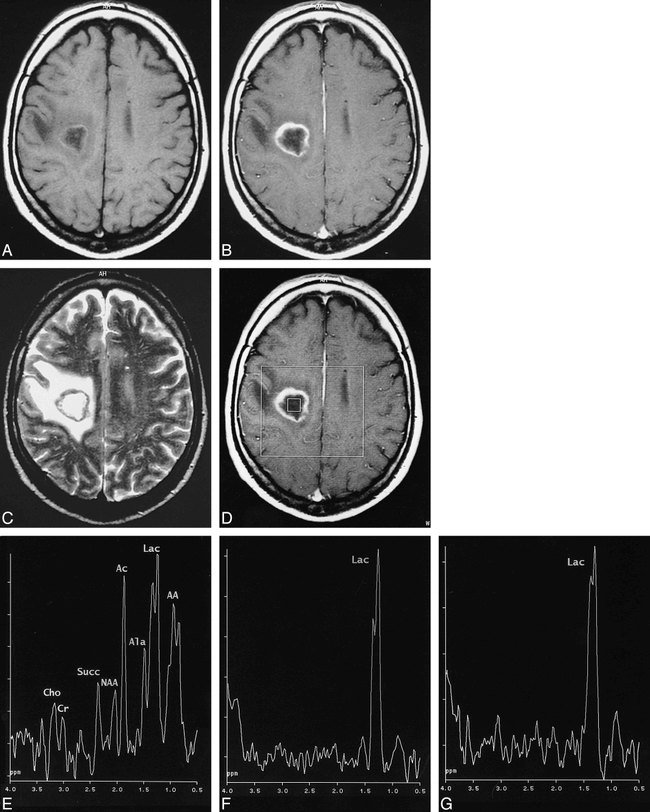
Patient 3
A 54-year-old man with a history of vertigo had sudden onset of left upper extremity weakness. He had no fever, and was admitted to the hospital 1 day after symptom onset. CT on days 2 and 4 showed a single, small, cystic, contrast-enhancing lesion in the right parietal lobe, 1.5 cm in size initially, increasing to 3 cm on day 4. MR imaging on day 6 (Fig 3A–D) showed no further increase in size. MR spectroscopy performed at the same time (Fig 3E) revealed a similar spectral pattern as in patient 2, with succinate, acetate, alanine, lactate, and amino acid resonances (Table 1). High-dose antibiotic therapy was started immediately (day 6). A follow-up examination (day 20) of this patient revealed a dramatic change in the spectrum from the abscess cyst. All resonances but lactate disappeared (Fig 3F) and this finding was confirmed by two more follow-up examinations on days 29 (Fig 3G) and 113, respectively. Bacteria responsible for the abscess could not be traced until day 35, when a CSF sample was positive for Streptococcus mitis and Enterococcus faecalis. During antibiotic therapy, the lesion size decreased very slowly to 1.5 cm at day 113, when neurosurgery was performed. Histopathologic findings confirmed the diagnosis of abscess.

MR images of patient 3.
A–C, T1-weighted axial spin-echo images (630/17/2) before (A) and after (B) contrast injection, and T2-weighted axial fast spin-echo image (3710/90/1) (C).
D, Image shows the area of the spectroscopy measurement and the placement of the voxel representing the spectrum (see E).
E, The proton spectrum (2D chemical-shift PRESS sequence, 1500/270/2) from the abscess cyst shows resonances representing succinate (Succ), acetate (Ac), alanine (Ala), lactate (Lac), and amino acids (AA). Visible small resonances of Cho, Cr, and NAA were interpreted to be caused by partial volume effects. A follow-up examination 2 weeks after the first MR spectroscopic measurement and the institution of high-dose antibiotic therapy revealed a dramatic change in the spectrum from the abscess cyst.
F and G, All resonances but lactate disappeared (F), a finding that was confirmed by another two follow-up examinations 9 days (G) and 84 days later.
The spectral changes specific for bacterial abscess seen in these cases of intracranial abscesses were restricted to the cystic part of the mass lesions. In the periphery of the cystic lesions, where edema was present, a decrease of the NAA/Cho ratio to values of 1.1 (Cho/Cr = 1) and 1.2 (Cho/Cr = 0.9) could be seen in patients 1 and 3, respectively. These decreases were interpreted to be caused by edema, as they normalized to values of >1.9 when the edema had subsided. In patient 2, the measurement area did not cover enough tissue with edema to allow calculation of the metabolic ratio.
Discussion
MR spectroscopy is a potential tool for noninvasive evaluation of cystic brain lesions in which there is a differential diagnosis between brain abscess and noninfectious ring-enhancing brain lesions, such as primary brain tumors, lymphomas, brain metastases, and tuberculomas because of specific changes in MR spectra from abscess cavities. Furthermore, the fact that these spectral changes disappear with effective antibiotic treatment, as shown in our study, underlines the necessity that the neuroradiologist or spectroscopist interpreting the spectra be aware of the case history and that the MR spectroscopic examinations are performed as early as possible, preferably before the start of high-dose antibiotic therapy. Knowledge of the expected spectral changes after treatment also offers the possibility of evaluating the efficacy of nonsurgical treatment of brain abscesses.
The specific changes in MR spectra from abscess cavities, which are illustrated in patients 2 and 3, include the occurrence of resonances representing 1) acetate, lactate, pyruvate, and succinate, which are known metabolic end-products arising from microorganisms (except for lactate, which is a nonspecific marker in many human diseases, these metabolites have not been reported in human brain tissue other than in conjunction with infection) and 2) amino-acid signals at 0.9 ppm, seen in many brain abscesses and assigned to valine, leucine, and isoleucine (considered to represent accumulated end-products of proteolysis caused by proteolytic enzymes secreted by microorganisms, or polymorphonuclear leukocytes in pus, or both.) (5, 6, 8, 17). To our knowledge, no MR spectra similar to those presented in this or the above-mentioned studies have been reported to represent brain lesions other than bacterial infections or cysticercosis (14). Although the earlier mentioned metabolites represent well-known end-products of protein and carbohydrate metabolism of various bacterial strains causing abscesses, it is still unclear whether MR spectroscopy can be used to identify the various bacterial strains present in abscesses, which is usually done by means of gas-liquid chromatography (8).
The literature published before April 1998 contains reports of 67 cases of intracranial abscesses, with a total of 70 lesions examined with in vivo proton MR spectroscopy (2, 4–16). Nineteen of these cases with a single brain abscess might have been published twice. For 31 of these lesions, additional in vitro MR spectroscopic examinations were performed. All authors concluded that MR spectroscopy might be of additional value to morphologic methods in the diagnosis of brain abscess. The MR spectroscopic results in our patient 2, who had been treated with antibiotics for only 1 day, and in our patient 3, who was examined by MR spectroscopy before the start of therapy, confirm the specific spectral pattern of brain abscess reported previously. Furthermore, the absence of all specific abscess metabolites in patient 1, who had been treated with antibiotics 40 days before the first MR spectroscopic examination, and the dramatic spectral changes observed under antibiotic treatment in patient 3, show that the spectral specificity is only valid for untreated abscesses or soon after the start of treatment. The finding of a single lactate peak after treatment in patients 1 and 3 was nonspecific and can be seen in several other conditions, such as cystic gliomas or metastases, which have a similar morphologic appearance as brain abscess. Our material is small but is supported by a recently published study by Dev et al (4). Further prospective studies with a larger number of cases will be necessary to evaluate thoroughly the particular spectral peaks and the time course of the spectral changes seen with antibiotic treatment.
Acknowledgments
We express our gratitude to our colleagues from the Departments of Diagnostic Radiology, Neurosurgery, Neurology, and Pathology, University Hospital Lund, Sweden.
Footnotes
↵1 Supported by grants from the Swedish Medical Research Council, the Swedish Cancer Society, and the Children Cancer Foundation of Sweden.
↵2 Presented in part at the annual meetings of the International Society for Magnetic Resonance in Medicine, Sydney, 1998, and the European Congress in Neuroradiology, Lisbon 1998.
↵3 Address reprint requests to Isabella M. Burtscher, MD.
References
- Received July 31, 1998.
- Copyright © American Society of Neuroradiology
In this issue


Jump to section
Related Articles
Cited By...
- Multiparametric imaging in the evaluation of intracerebral abscesses
- Multiparametric imaging in the evaluation of intracerebral abscesses
- Intracranial hydatid cyst: imaging findings of a rare disease
- Role of imaging in the diagnosis of acute bacterial meningitis and its complications
- In Vivo Proton MR Spectroscopy Evaluation of Pyogenic Brain Abscesses: A Report of 194 Cases