
Article Figures & Data
Figures
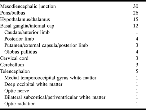
- fig 1.
A, Coronal T2-weighted image (4000/90/2 [TR/TE/excitations]) at the level of the crus cerebri nicely shows heterogeneous left MDJ lesion with extensive edema, sparing the red nucleus.
B, Coronal noncontrast T1-weighted image (300/15/3) at the same level as A reveals a hemorrhagic focus in the lesion.
C, Coronal T2-weighted image (4000/90/2) posterior to A shows extension of perilesional edema caudally to the superior cerebellar peduncle and pontine tegmentum, and upward to the white matter of the temporal lobe, external capsule, and thalamus.
D, Caudal extension of the edema toward the pontine tegmentum is seen on axial T2-weighted image (4000/90/1).
E, Two years later, after another relapse of the disease, coronal T2-weighted image (4000/90/1) reveals a contralateral MDJ lesion. The left-sided lesion now has shrunk to a small hypointense area.
F, Contrast-enhanced T1-weighted image (660/17/1) shows enhancement of the new right MDJ lesion.
G and H, Similar extension of edema as observed in C and D is seen in the right mesencephalopontine region of the brain stem on T2-weighted images (4000/90/1).
- fig 2.
Axial T2-weighted image (2400/110/2) shows inhomogeneous hyperintense lesion located in the mid pons.
fig 3. Axial T2-weighted image (2400/110/2) at the pontine level shows a right-sided pontine lesion that does not cross the midline.
- fig 4.
A and B, Axial (A) and coronal (B) T2-weighted images (4000/90/2) reveal hyperintense lesions bilaterally in the middle cerebellar peduncles and deep cerebellar white matter
- fig 5.
A–C, Midsagittal cervical lateral T2-weighted image (2200/80/1) (A) and axial T2-weighted image (4000/90/2) through the cervical medullary junction (B) show posteriorly located paracentral hyperintense lesion with mild cervicomedullary enlargement. The pattern of extension up to the inferior cerebellar peduncle suggests involvement of the dorsal columns. Midsagittal cervical lateral T1-weighted image (300/15/3) (C) shows slight enlargement of cervical cord
- fig 6.
A, Axial T2-weighted image (3500/90/2) shows a well-defined deep right occipital white matter lesion (asterisk) and a subcortical linear hyperintensity (arrow).
B, Coronal T2-weighted image (3500/90/1) in a different patient shows multiple subcortical white matter and right MDJ and pontine lesions (asterisk and arrowheads).
- fig 7.
A, T2-weighted image (4000/90/2) shows chronic left MDJ lesion (double arrowhead) and ipsilateral lenticulostriatal lesions (arrow).
B, Wallerian degeneration of the optic radiation is evident on paraatrial section of the same sequence (double arrowheads).
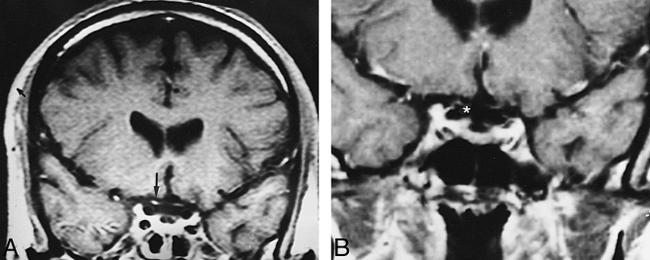
- fig 8.
A, Contrast-enhanced coronal T1-weighted image (600/15/1) shows bilateral optic nerve atrophy and marked enhancement of the prechiasmatic segment of the right optic nerve (arrow).
B, Contrast enhancement of the nerve disappeared after treatment (asterisk).
- fig 9.
A, T2-weighted image (2400/110/2) shows round hyperintense lesion in the posterior and middle third of the right corona radiata (asterisk) associated with rather poorly defined periatrial T2 hyperintensities (arrowheads).
B, Coronal T2-weighted image (4000/90/2) shows signal intensity changes along the right corticospinal tract, representative of secondary wallerian degeneration (double arrowheads), along with basal ganglia and pontine lesions (asterisks).
- fig 10.
Histologic section of NBS shows a totally thrombosed medium-sized venous vessel. Focal fibrinoid necrosis (arrowheads) and moderate amount of lymphocyte and plasma cell infiltration is visible in the vessel wall. At the right side of the vessel, necrotic brain tissue with some newly thrombosed small vessel and mononuclear inflammatory infiltration (mainly lymphocytes and histiocytes) is seen (arrows). On the left, there is severe astrogliosis with some gemistocytic differentiation (H and E, original magnification ×100).
fig 11. Schematic representation of the intraaxial venous system of normal parenchyma. Supratentorially, a medullary vein (1) permits bidirectional flow. In the brain stem, especially at the mesencephalic level, intraaxial veno-venous anastomosis (2) is sparse and venous flow is centrifugal, toward the pial veins (pv). ev indicates ependymal vein.


Tables
 Table 1:
Lesion distribution in 65 patients with neuro-Beh;alcet syndrome


In this issue


Jump to section
Related Articles
Cited By...
- Neurological involvement by Behcets syndrome: clinical features, diagnosis, treatment and outcome
- Case of elderly onset possible neuro-Behcets disease with HLA-B51 homozygosity
- Clinical Reasoning: Rare Cause of Hemiparesis and Ataxia in a 36-Year-Old Man
- Neuro-Behcet's disease mimicking a cranial tumour
- Clinical characteristics of pediatric-onset neuro-Behcet disease
- Evaluation of Parenchymal Neuro-Behcet Disease by Using Susceptibility-Weighted Imaging
- Association between sarcoidosis and Behcet disease in a young woman with symptoms mimicking myasthaenia gravis
- Primary CNS vasculitis with spinal cord involvement
- An unusual case of hemiparesis in a young woman
- Bilateral subdural effusion in a patient with neuro-Behcet's disease
- Brain Perfusion SPECT in Juvenile Neuro-Behcet's Disease