
Article Figures & Data
Figures
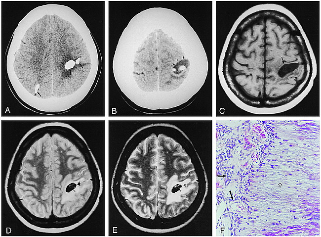
- fig 1.
Case 1: 47-year-old woman.
A and B, Noncontrast CT scans show multiple round calcifications (solid arrows) and eccentric cysts (arrowheads) with moderate edema in the left frontoparietal (open arrow) and right parietal lobes.
C, On T1-weighted MR image (420/14/2 [TR/TE/excitations]), the cyst (arrow) is isointense with CSF; subtle, mottled dark signal intensity is also noted.
D and E, On proton density–weighted (2600/22/2) (D) and T2-weighted (2600/90/2) (E) images, the lesions have high signal intensity with central areas of low signal intensity (arrows).
F, Histopathologic specimen shows proliferating blood vessels surrounded by meningothelial cells (closed arrows) and fibrillary calcification (open arrow) (hematoxylin-eosin, original magnification ×100). This specimen was obtained from the cortex of the left parietal lobe.
- fig 2.
Case 2: 53-year-old man.
A, Noncontrast CT scan reveals multiple round calcifications (closed arrows) with eccentric cysts (open arrow) and moderate edema.
B, On T1-weighted image (420/14/2), the lesions show inhomogeneous hypo- and intermediate signal intensity in the left frontal and parietal lobes.
C, On T2-weighted image (2600/90/2), areas of heterogeneous signal intensity were noted in the left frontal and parietal lobes, caused by calcification (closed arrows), cyst (open arrow), and edema.
D and E, On contrast-enhanced T1-weighted image (420/14/2), the lesions show irregular enhancement (arrows).


